ADP-2Ho as a phasing tool for nucleotide-containing proteins.
Ku, S.Y., Smith, G.D., Howell, P.L.(2007) Acta Crystallogr D Biol Crystallogr 63: 493-499
- PubMed: 17372354 Search on PubMed
- DOI: https://doi.org/10.1107/S0907444907006592
- Primary Citation Related Structures:
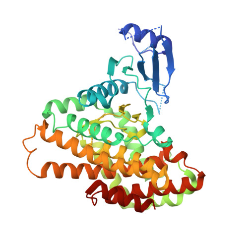
2OLC - PubMed Abstract:
Trivalent holmium ions were shown to isomorphously replace magnesium ions to form an ADP-2Ho complex in the nucleotide-binding domain of Bacillus subtilis 5-methylthioribose (MTR) kinase. This nucleotide-holmium complex provided sufficient phasing power to allow SAD and SIRAS phasing of this previously unknown structure using the L(III) absorption edge of holmium. The structure of ADP-2Ho reveals that the two Ho ions are approximately 4 A apart and are likely to share their ligands: the phosphoryl O atoms of ADP and a water molecule. The structure determination of MTR kinase using data collected using Cu Kalpha X-radiation was also attempted. Although the heavy-atom substructure determination was successful, interpretation of the map was more challenging. The isomorphous substitution of holmium for magnesium in the MTR kinase-nucleotide complex suggests that this could be a useful phasing tool for other metal-dependent nucleotide-containing proteins.
- Program in Molecular Structure and Function, Research Institute, Hospital for Sick Children, 555 University Avenue, Toronto, Ontario M5G 1X8, Canada.
Organizational Affiliation: