2OJT | pdb_00002ojt

Structure and mechanism of kainate receptor modulation by anions


Experimental Data Snapshot

  • Method: X-RAY DIFFRACTION
  • Resolution: 1.95 Å
  • R-Value Free: 
    0.233 (Depositor), 0.234 (DCC) 
  • R-Value Work: 
    0.190 (Depositor), 0.207 (DCC) 
  • R-Value Observed: 
    0.192 (Depositor) 

Starting Model: experimental
View more details

wwPDB Validation 3D Report Full Report

Validation slider image for 2OJT

Ligand Structure Quality Assessment 


This is version 1.5 of the entry. See complete history

Literature

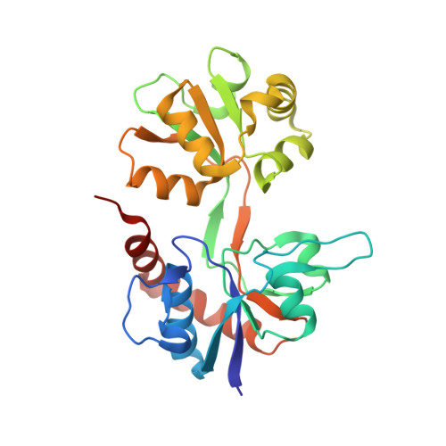
Structure and mechanism of kainate receptor modulation by anions.

Plested, A.J.Mayer, M.L.

(2007) Neuron 53: 829-841

  • DOI: https://doi.org/10.1016/j.neuron.2007.02.025
  • Primary Citation Related Structures: 
    2OJT

  • PubMed Abstract: 

    L-glutamate, the major excitatory neurotransmitter in the human brain, activates a family of ligand-gated ion channels, the major subtypes of which are named AMPA, kainate, and NMDA receptors. In common with many signal transduction proteins, glutamate receptors are modulated by ions and small molecules, including Ca(2+), Mg(2+), Zn(2+), protons, polyamines, and steroids. Strikingly, the activation of kainate receptors by glutamate requires the presence of both Na(+) and Cl(-) in the extracellular solution, and in the absence of these ions, receptor activity is abolished. Here, we identify the site and mechanism of action of anions. Surprisingly, we find that Cl(-) ions are essential structural components of kainate receptors. Cl(-) ions bind in a cavity formed at the interface between subunits in a dimer pair. In the absence of Cl(-), dimer stability is reduced, the rate of desensitization increases, and the fraction of receptors competent for activation by glutamate drops precipitously.


  • Organizational Affiliation
    • Laboratory of Cellular and Molecular Neurophysiology, Porter Neuroscience Research Center, National Institute of Child Health and Human Development, National Institutes of Health, Department of Health and Human Services, Bethesda, MD 20892, USA.

Macromolecule Content 

  • Total Structure Weight: 59.77 kDa 
  • Atom Count: 4,706 
  • Modeled Residue Count: 502 
  • Deposited Residue Count: 516 
  • Unique protein chains: 1

Macromolecules

Find similar proteins by:|  3D Structure
Entity ID: 1
MoleculeChains  Sequence LengthOrganismDetailsImage
Glutamate receptor, ionotropic kainate 1
A, B
258Rattus norvegicusMutation(s): 1 
Gene Names: Grik1Glur5
UniProt
Find proteins for P22756 (Rattus norvegicus)
Explore P22756 
Go to UniProtKB:  P22756
Entity Groups
Sequence Clusters30% Identity50% Identity70% Identity90% Identity95% Identity100% Identity
UniProt GroupP22756
Sequence Annotations
Expand
Reference Sequence

Small Molecules

Ligands 3 Unique
IDChains Name / Formula / InChI Key2D Diagram3D Interactions
UBA

Query on UBA


Download:Ideal Coordinates CCD File
E [auth A],
H [auth B]
(S)-1-(2-AMINO-2-CARBOXYETHYL)-3(2-CARBOXYTHIOPHENE-3-YL-METHYL)-5-METHYLPYRIMIDINE-2,4-DIONE
C14 H15 N3 O6 S
ZTAZUCRXCRXNSU-VIFPVBQESA-N
1PE

Query on 1PE


Download:Ideal Coordinates CCD File
D [auth A],
G [auth B]
PENTAETHYLENE GLYCOL
C10 H22 O6
JLFNLZLINWHATN-UHFFFAOYSA-N
BR

Query on BR


Download:Ideal Coordinates CCD File
C [auth A],
F [auth B]
BROMIDE ION
Br
CPELXLSAUQHCOX-UHFFFAOYSA-M

Experimental Data & Validation

Experimental Data

  • Method: X-RAY DIFFRACTION
  • Resolution: 1.95 Å
  • R-Value Free:  0.233 (Depositor), 0.234 (DCC) 
  • R-Value Work:  0.190 (Depositor), 0.207 (DCC) 
  • R-Value Observed: 0.192 (Depositor) 
Space Group: C 2 2 21
Unit Cell:
Length ( Å )Angle ( ˚ )
a = 97.598α = 90
b = 97.732β = 90
c = 128.688γ = 90
Software Package:
Software NamePurpose
REFMACrefinement
HKL-2000data collection
HKL-2000data reduction
HKL-2000data scaling
AMoREphasing

Structure Validation

View Full Validation Report


Ligand Structure Quality Assessment 


Entry History 

Deposition Data

  • Released Date: 2007-04-03 
  • Deposition Author(s): Mayer, M.L.

Revision History  (Full details and data files)

  • Version 1.0: 2007-04-03
    Type: Initial release
  • Version 1.1: 2008-05-01
    Changes: Version format compliance
  • Version 1.2: 2011-07-13
    Changes: Advisory, Refinement description, Version format compliance
  • Version 1.3: 2017-08-02
    Changes: Source and taxonomy
  • Version 1.4: 2021-10-20
    Changes: Database references, Derived calculations
  • Version 1.5: 2023-08-30
    Changes: Data collection, Refinement description