2OIT | pdb_00002oit

Crystal Structure of the N-terminal Domain of the Human Proto-oncogene Nup214/CAN


Experimental Data Snapshot

  • Method: X-RAY DIFFRACTION
  • Resolution: 1.65 Å
  • R-Value Free: 
    0.237 (Depositor), 0.243 (DCC) 
  • R-Value Work: 
    0.199 (Depositor), 0.205 (DCC) 

wwPDB Validation 3D Report Full Report

Validation slider image for 2OIT

This is version 1.4 of the entry. See complete history

Literature

Crystal structure of the N-terminal domain of the human protooncogene Nup214/CAN.

Napetschnig, J.Blobel, G.Hoelz, A.

(2007) Proc Natl Acad Sci U S A 104: 1783-1788

  • DOI: https://doi.org/10.1073/pnas.0610828104
  • Primary Citation Related Structures: 
    2OIT

  • PubMed Abstract: 

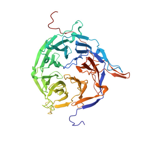
    The mammalian nuclear pore complex (NPC) is an approximately 120-MDa proteinaceous assembly consisting of approximately 30 proteins and is the sole gate in the nuclear envelope. The human protooncogene Nup214 was first identified as a target for chromosomal translocation involved in leukemogenesis. Nup214 is located on the cytoplasmic face of the NPC and is implicated in anchoring the cytoplasmic filaments of the NPC and recruiting the RNA helicase Ddx19. Here, we present the crystal structure of the human Nup214 N-terminal domain at 1.65-A resolution. The structure reveals a seven-bladed beta-propeller followed by a 30-residue C-terminal extended peptide segment, which folds back onto the beta-propeller and binds to its bottom face. The beta-propeller repeats lack any recognizable sequence motif and are distinguished by extensive insertions between the canonical beta-strands. We propose a mechanism by which the C-terminal peptide extension is involved in NPC assembly.


  • Organizational Affiliation
    • Laboratory of Cell Biology and Howard Hughes Medical Institute, The Rockefeller University, New York, NY 10021, USA.

Macromolecule Content 

  • Total Structure Weight: 48.31 kDa 
  • Atom Count: 3,849 
  • Modeled Residue Count: 434 
  • Deposited Residue Count: 434 
  • Unique protein chains: 1

Macromolecules

Find similar proteins by:|  3D Structure
Entity ID: 1
MoleculeChains  Sequence LengthOrganismDetailsImage
Nucleoporin 214kDa434Homo sapiensMutation(s): 0 
Gene Names: NUP214
UniProt & NIH Common Fund Data Resources
Find proteins for P35658 (Homo sapiens)
Explore P35658 
Go to UniProtKB:  P35658
PHAROS:  P35658
GTEx:  ENSG00000126883 
Entity Groups
Sequence Clusters30% Identity50% Identity70% Identity90% Identity95% Identity100% Identity
UniProt GroupP35658
Sequence Annotations
Expand
Reference Sequence

Small Molecules

Ligands 1 Unique
IDChains Name / Formula / InChI Key2D Diagram3D Interactions
MES

Query on MES


Download:Ideal Coordinates CCD File
B [auth A]2-(N-MORPHOLINO)-ETHANESULFONIC ACID
C6 H13 N O4 S
SXGZJKUKBWWHRA-UHFFFAOYSA-N

Experimental Data & Validation

Experimental Data

  • Method: X-RAY DIFFRACTION
  • Resolution: 1.65 Å
  • R-Value Free:  0.237 (Depositor), 0.243 (DCC) 
  • R-Value Work:  0.199 (Depositor), 0.205 (DCC) 
Space Group: P 21 21 21
Unit Cell:
Length ( Å )Angle ( ˚ )
a = 52.381α = 90
b = 81.14β = 90
c = 102.607γ = 90
Software Package:
Software NamePurpose
DENZOdata reduction
SCALEPACKdata scaling
CNSrefinement
PDB_EXTRACTdata extraction
HKL-2000data collection
HKL-2000data reduction
SHARPphasing

Structure Validation

View Full Validation Report


Entry History 

Deposition Data

Revision History  (Full details and data files)

  • Version 1.0: 2007-02-06
    Type: Initial release
  • Version 1.1: 2008-05-01
    Changes: Version format compliance
  • Version 1.2: 2011-07-13
    Changes: Version format compliance
  • Version 1.3: 2017-10-18
    Changes: Refinement description
  • Version 1.4: 2023-12-27
    Changes: Data collection, Database references, Derived calculations