Structure-based mutagenesis of the substrate-recognition domain of Nrdp1/FLRF identifies the binding site for the receptor tyrosine kinase ErbB3.
Bouyain, S., Leahy, D.J.(2007) Protein Sci 16: 654-661
- PubMed: 17384230 Search on PubMedSearch on PubMed Central
- DOI: https://doi.org/10.1110/ps.062700307
- Primary Citation Related Structures:
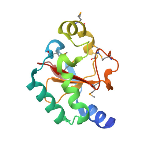
2OGB - PubMed Abstract:
The E3 ubiquitin ligase neuregulin receptor degrading protein 1 (Nrdp1) mediates the ligand-independent degradation of the epidermal growth factor receptor family member ErbB3/HER3. By regulating cellular levels of ErbB3, Nrdp1 influences ErbB3-mediated signaling, which is essential for normal vertebrate development. Nrdp1 belongs to the tripartite or RBCC (RING, B-box, coiled-coil) family of ubiquitin ligases in which the RING domain is responsible for ubiquitin ligation and a variable C-terminal region mediates substrate recognition. We report here the 1.95 A crystal structure of the C-terminal domain of Nrdp1 and show that this domain is sufficient to mediate ErbB3 binding. Furthermore, we have used site-directed mutagenesis to map regions of the Nrdp1 surface that are important for interacting with ErbB3 and mediating its degradation in transfected cells. The ErbB3-binding site localizes to a region of Nrdp1 that is conserved from invertebrates to vertebrates, in contrast to ErbB3, which is only found in vertebrates. This observation suggests that Nrdp1 uses a common binding site to recognize its targets in different species.
- Department of Biophysics and Biophysical Chemistry, The Johns Hopkins University School of Medicine, Baltimore, Maryland 21205, USA.
Organizational Affiliation: