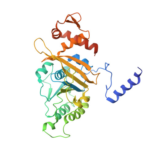
Snapshots of RecA protein involving movement of the C-domain and different conformations of the DNA-binding loops: crystallographic and comparative analysis of 11 structures of Mycobacterium smegmatis RecA
Krishna, R., Prabu, J.R., Manjunath, G.P., Datta, S., Chandra, N.R., Muniyappa, K., Vijayan, M.(2007) J Mol Biology 367: 1130-1144
- PubMed: 17306300
- DOI: https://doi.org/10.1016/j.jmb.2007.01.058
- Primary Citation of Related Structures:
2ODN, 2ODW, 2OE2, 2OEP, 2OES, 2OFO - PubMed Abstract:
Mycobacterium smegmatis RecA and its nucleotide complexes crystallize in three different, but closely related, forms characterized by specific ranges of unit cell dimensions. The six crystals reported here and five reported earlier, all grown under the same or very similar conditions, belong to these three forms, all in space group P6(1). They include one obtained by reducing relative humidity around the crystal. In all crystals, RecA monomers form filaments around a 6(1) screw axis. Thus, the c-dimension of the crystal corresponds to the pitch of the RecA filament. As reported for Escherichia coli RecA, the variation in the pitch among the three forms correlates well with the motion of the C-terminal domain of the RecA monomers with respect to the main domain. The domain motion is compatible with formation of inactive as well as active RecA filaments involving monomers with a fully ordered C domain. It does not appear to influence the movement upon nucleotide-binding of the switch residue, which is believed to provide the trigger for transmitting the effect of nucleotide binding to the DNA-binding region. Interestingly, partial dehydration of the crystal results in the movement of the residue similar to that caused by nucleotide binding. The ordering of the DNA-binding loops, which present ensembles of conformations, is also unaffected by domain motion. The conformation of loop L2 appears to depend upon nucleotide binding, presumably on account of the movement of the switch residue that forms part of the loop. The conformations of loops L1 and L2 are correlated and have implications for intermolecular communications within the RecA filament. The structures resulting from different orientations of the C domain and different conformations of the DNA-binding loops appear to represent snapshots of the RecA at different phases of activity, and provide insights into the mechanism of action of RecA.
- Molecular Biophysics Unit, Indian Institute of Science, Bangalore, 560 012, India.
Organizational Affiliation:


