2OBL | pdb_00002obl

Structural and biochemical analysis of a prototypical ATPase from the type III secretion system of pathogenic bacteria


Experimental Data Snapshot

  • Method: X-RAY DIFFRACTION
  • Resolution: 1.80 Å
  • R-Value Free: 
    0.232 (Depositor), 0.230 (DCC) 
  • R-Value Work: 
    0.195 (Depositor), 0.195 (DCC) 
  • R-Value Observed: 
    0.197 (Depositor) 

wwPDB Validation 3D Report Full Report

Validation slider image for 2OBL

This is version 1.4 of the entry. See complete history

Literature

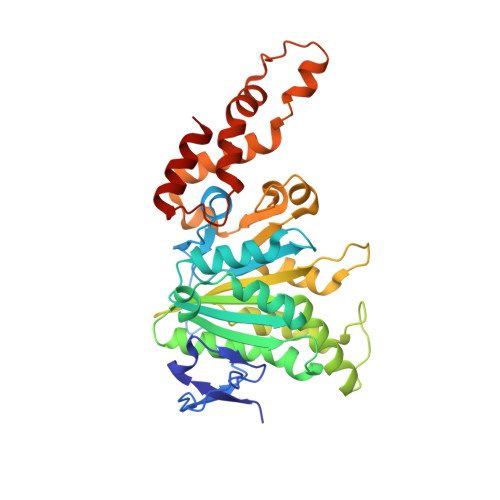
Structural analysis of a prototypical ATPase from the type III secretion system.

Zarivach, R.Vuckovic, M.Deng, W.Finlay, B.B.Strynadka, N.C.

(2007) Nat Struct Mol Biol 14: 131-137

  • DOI: https://doi.org/10.1038/nsmb1196
  • Primary Citation Related Structures: 
    2OBL, 2OBM

  • PubMed Abstract: 

    The type III secretion system (T3SS) ATPase is the conserved and essential inner-membrane component involved in the initial stages of selective secretion of specialized T3SS virulence effector proteins from the bacterial cytoplasm through to the infected host cell, a process crucial to subsequent pathogenicity. Here we present the 1.8-A-resolution crystal structure of the catalytic domain of the prototypical T3SS ATPase EscN from enteropathogenic Escherichia coli (EPEC). Along with in vitro and in vivo mutational analysis, our data show that the T3SS ATPases share similarity with the F1 ATPases but have important structural and sequence differences that dictate their unique secretory role. We also show that T3SS ATPase activity is dependent on EscN oligomerization and describe the molecular features and possible functional implications of a hexameric ring model.


  • Organizational Affiliation
    • Department of Biochemistry and Molecular Biology and the Center for Blood Research, University of British Columbia, 2350 Health Sciences Mall, Vancouver, British Columbia V6T 1Z3, Canada.

Macromolecule Content 

  • Total Structure Weight: 37.84 kDa 
  • Atom Count: 2,888 
  • Modeled Residue Count: 346 
  • Deposited Residue Count: 347 
  • Unique protein chains: 1

Macromolecules

Find similar proteins by:|  3D Structure
Entity ID: 1
MoleculeChains  Sequence LengthOrganismDetailsImage
EscN347Escherichia coli O127:H6 str. E2348/69Mutation(s): 1 
Gene Names: escN
EC: 7.4.2.8
UniProt
Find proteins for B7UMA6 (Escherichia coli O127:H6 (strain E2348/69 / EPEC))
Explore B7UMA6 
Go to UniProtKB:  B7UMA6
Entity Groups
Sequence Clusters30% Identity50% Identity70% Identity90% Identity95% Identity100% Identity
UniProt GroupB7UMA6
Sequence Annotations
Expand
Reference Sequence

Experimental Data & Validation

Experimental Data

  • Method: X-RAY DIFFRACTION
  • Resolution: 1.80 Å
  • R-Value Free:  0.232 (Depositor), 0.230 (DCC) 
  • R-Value Work:  0.195 (Depositor), 0.195 (DCC) 
  • R-Value Observed: 0.197 (Depositor) 
Space Group: P 21 21 2
Unit Cell:
Length ( Å )Angle ( ˚ )
a = 84.382α = 90
b = 77.609β = 90
c = 52.901γ = 90
Software Package:
Software NamePurpose
REFMACrefinement
ADSCdata collection
HKL-2000data reduction
HKL-2000data scaling
SHELXSphasing

Structure Validation

View Full Validation Report


Entry History 

Deposition Data

Revision History  (Full details and data files)

  • Version 1.0: 2007-01-30
    Type: Initial release
  • Version 1.1: 2008-05-01
    Changes: Version format compliance
  • Version 1.2: 2011-07-13
    Changes: Advisory, Source and taxonomy, Version format compliance
  • Version 1.3: 2021-10-20
    Changes: Database references, Derived calculations
  • Version 1.4: 2023-12-27
    Changes: Data collection