Structural basis of aquaporin inhibition by mercury.
Savage, D.F., Stroud, R.M.(2007) J Mol Biology 368: 607-617
- PubMed: 17376483 Search on PubMedSearch on PubMed Central
- DOI: https://doi.org/10.1016/j.jmb.2007.02.070
- Primary Citation Related Structures:
2O9D, 2O9E, 2O9F, 2O9G - PubMed Abstract:
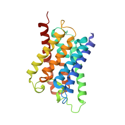
The aquaporin family of channels was defined based on the inhibition of water transport by mercurial compounds. Despite the important role of mercurials, little is known about the structural changes involved upon mercury binding leading to channel inhibition. To elucidate the mechanism we designed a mutant, T183C, of aquaporin Z (AqpZ) patterned after the known mercury-sensitive site of aquaporin 1 (AQP1) and determined the X-ray crystal structures of the unbound and mercury blocked states. Superposition of the two structures shows no conformational rearrangement upon mercury binding. In the blocked structure, there are two mercury sites, one bound to Cys183 and occluding the pore, and a second, also bound to the same cysteine but found buried in an interstitial cavity. To test the mechanism of blockade we designed a different mutant, L170C, to produce a more effective mercury block at the pore site. In a dose-response inhibition study, this mutant was 20 times more sensitive to mercury than wild-type AqpZ and four times more sensitive than T183C. The X-ray structure of L170C shows four mercury atoms at, or near, the pore site defined in the T183C structure and no structural change upon mercury binding. Thus, we elucidate a steric inhibition mechanism for this important class of channels by mercury.
- Graduate Group in Biophysics, University of California at San Francisco, San Francisco, CA 94158, USA. savage@msg.ucsf.edu
Organizational Affiliation: