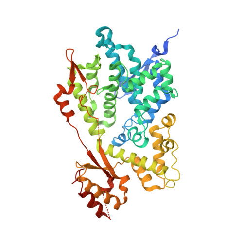
2O6I

Structure of an Enterococcus Faecalis HD Domain Phosphohydrolase


Experimental Data Snapshot

  • Method: X-RAY DIFFRACTION
  • Resolution: 2.55 Å
  • R-Value Free: 0.317 
  • R-Value Work: 0.248 
  • R-Value Observed: 0.252 

wwPDB Validation   3D Report Full Report


Ligand Structure Quality Assessment 


This is version 1.4 of the entry. See complete history


Literature

Characterization of the deoxynucleotide triphosphate triphosphohydrolase (dNTPase) activity of the EF1143 protein from Enterococcus faecalis and crystal structure of the activator-substrate complex.

Vorontsov, I.I.Minasov, G.Kiryukhina, O.Brunzelle, J.S.Shuvalova, L.Anderson, W.F.

(2011) J Biol Chem 286: 33158-33166

  • DOI: 10.1074/jbc.M111.250456
  • Primary Citation of Related Structures:  
    2O6I, 3IRH

  • PubMed Abstract: 
  • The EF1143 protein from Enterococcus faecalis is a distant homolog of deoxynucleotide triphosphate triphosphohydrolases (dNTPases) from Escherichia coli and Thermus thermophilus. These dNTPases are important components in the regulation of the dNTP pool in bacteria ...

    The EF1143 protein from Enterococcus faecalis is a distant homolog of deoxynucleotide triphosphate triphosphohydrolases (dNTPases) from Escherichia coli and Thermus thermophilus. These dNTPases are important components in the regulation of the dNTP pool in bacteria. Biochemical assays of the EF1143 dNTPase activity demonstrated nonspecific hydrolysis of all canonical dNTPs in the presence of Mn(2+). In contrast, with Mg(2+) hydrolysis required the presence of dGTP as an effector, activating the degradation of dATP and dCTP with dGTP also being consumed in the reaction with dATP. The crystal structure of EF1143 and dynamic light scattering measurements in solution revealed a tetrameric oligomer as the most probable biologically active unit. The tetramer contains four dGTP specific allosteric regulatory sites and four active sites. Examination of the active site with the dATP substrate suggests an in-line nucleophilic attack on the α-phosphate center as a possible mechanism of the hydrolysis and two highly conserved residues, His-129 and Glu-122, as an acid-base catalytic dyad. Structural differences between EF1143 apo and holo forms revealed mobility of the α3 helix that can regulate the size of the active site binding pocket and could be stabilized in the open conformation upon formation of the tetramer and dGTP effector binding.


    Organizational Affiliation

    Department of Molecular Pharmacology and Biological Chemistry, Northwestern University Feinberg School of Medicine, Chicago, Illinois 60611, USA.


Macromolecules
Find similar proteins by:  (by identity cutoff)  |  3D Structure
Entity ID: 1
MoleculeChainsSequence LengthOrganismDetailsImage
HD domain proteinA, B480Enterococcus faecalis V583Mutation(s): 0 
Gene Names: EF-1143EF_1143
UniProt
Find proteins for Q836G9 (Enterococcus faecalis (strain ATCC 700802 / V583))
Explore Q836G9 
Go to UniProtKB:  Q836G9
Entity Groups  
Sequence Clusters30% Identity50% Identity70% Identity90% Identity95% Identity100% Identity
UniProt GroupQ836G9
Protein Feature View
Expand
  • Reference Sequence
Small Molecules
Ligands 3 Unique
IDChainsName / Formula / InChI Key2D Diagram3D Interactions
HED
Query on HED

Download Ideal Coordinates CCD File 
E [auth A]2-HYDROXYETHYL DISULFIDE
C4 H10 O2 S2
KYNFOMQIXZUKRK-UHFFFAOYSA-N
 Ligand Interaction
ZN
Query on ZN

Download Ideal Coordinates CCD File 
C [auth A],
F [auth B]
ZINC ION
Zn
PTFCDOFLOPIGGS-UHFFFAOYSA-N
 Ligand Interaction
CL
Query on CL

Download Ideal Coordinates CCD File 
D [auth A],
G [auth B]
CHLORIDE ION
Cl
VEXZGXHMUGYJMC-UHFFFAOYSA-M
 Ligand Interaction
Experimental Data & Validation

Experimental Data

  • Method: X-RAY DIFFRACTION
  • Resolution: 2.55 Å
  • R-Value Free: 0.317 
  • R-Value Work: 0.248 
  • R-Value Observed: 0.252 
  • Space Group: P 32 2 1
Unit Cell:
Length ( Å )Angle ( ˚ )
a = 109.913α = 90
b = 109.913β = 90
c = 182.412γ = 120
Software Package:
Software NamePurpose
REFMACrefinement
MAR345data collection
HKL-2000data reduction
HKL-2000data scaling
PHENIXphasing
SHARPphasing

Structure Validation

View Full Validation Report


Ligand Structure Quality Assessment 


Entry History 

Deposition Data

Revision History  (Full details and data files)

  • Version 1.0: 2006-12-19
    Type: Initial release
  • Version 1.1: 2008-05-01
    Changes: Version format compliance
  • Version 1.2: 2011-07-13
    Changes: Advisory, Source and taxonomy, Version format compliance
  • Version 1.3: 2011-11-23
    Changes: Database references
  • Version 1.4: 2017-10-18
    Changes: Refinement description