2O3Z | pdb_00002o3z

X-ray crystal structure of LpxC complexed with 3-heptyloxybenzoate


Experimental Data Snapshot

  • Method: X-RAY DIFFRACTION
  • Resolution: 2.25 Å
  • R-Value Free: 
    0.227 (Depositor), 0.213 (DCC) 
  • R-Value Work: 
    0.197 (Depositor), 0.190 (DCC) 

Starting Model: experimental
View more details

wwPDB Validation 3D Report Full Report

Validation slider image for 2O3Z

Ligand Structure Quality Assessment 


This is version 1.4 of the entry. See complete history

Literature

Amphipathic benzoic acid derivatives: synthesis and binding in the hydrophobic tunnel of the zinc deacetylase LpxC.

Shin, H.Gennadios, H.A.Whittington, D.A.Christianson, D.W.

(2007) Bioorg Med Chem 15: 2617-2623

  • DOI: https://doi.org/10.1016/j.bmc.2007.01.044
  • Primary Citation Related Structures: 
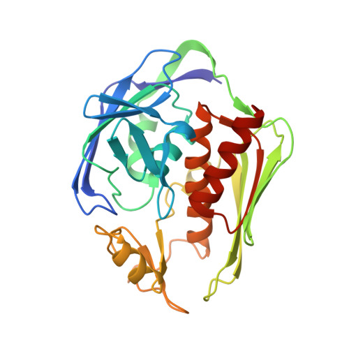
    2O3Z

  • PubMed Abstract: 

    The first committed step in lipid A biosynthesis is catalyzed by uridine diphosphate-(3-O-(R-3-hydroxymyristoyl))-N-acetylglucosamine deacetylase (LpxC), a zinc-dependent deacetylase, and inhibitors of LpxC may be useful in the development of antibacterial agents targeting a broad spectrum of Gram-negative bacteria. Here, we report the design of amphipathic benzoic acid derivatives that bind in the hydrophobic tunnel in the active site of LpxC. The hydrophobic tunnel accounts for the specificity of LpxC toward substrates and substrate analogues bearing a 3-O-myristoyl substituent. Simple benzoic acid derivatives bearing an aliphatic 'tail' bind in the hydrophobic tunnel with micromolar affinity despite the lack of a glucosamine ring like that of the substrate. However, although these benzoic acid derivatives each contain a negatively charged carboxylate 'warhead' intended to coordinate to the active site zinc ion, the 2.25A resolution X-ray crystal structure of LpxC complexed with 3-(heptyloxy)benzoate reveals 'backward' binding in the hydrophobic tunnel, such that the benzoate moiety does not coordinate to zinc. Instead, it binds at the outer end of the hydrophobic tunnel. Interestingly, these ligands bind with affinities comparable to those measured for more complicated substrate analogue inhibitors containing glucosamine ring analogues and hydroxamate 'warheads' that coordinate to the active site zinc ion. We conclude that the intermolecular interactions in the hydrophobic tunnel dominate enzyme affinity in this series of benzoic acid derivatives.


  • Organizational Affiliation
    • Department of Chemistry, University of San Francisco, 2130 Fulton Street, San Francisco, CA 94117-1080, USA.

Macromolecule Content 

  • Total Structure Weight: 63.01 kDa 
  • Atom Count: 4,554 
  • Modeled Residue Count: 534 
  • Deposited Residue Count: 542 
  • Unique protein chains: 1

Macromolecules

Find similar proteins by:|  3D Structure
Entity ID: 1
MoleculeChains  Sequence LengthOrganismDetailsImage
UDP-3-O-[3-hydroxymyristoyl] N-acetylglucosamine deacetylase
A, B
271Aquifex aeolicusMutation(s): 1 
Gene Names: lpxCenvA
EC: 3.5.1 (PDB Primary Data), 3.5.1.108 (UniProt)
UniProt
Find proteins for O67648 (Aquifex aeolicus (strain VF5))
Explore O67648 
Go to UniProtKB:  O67648
Entity Groups
Sequence Clusters30% Identity50% Identity70% Identity90% Identity95% Identity100% Identity
UniProt GroupO67648
Sequence Annotations
Expand
Reference Sequence

Small Molecules

Ligands 4 Unique
IDChains Name / Formula / InChI Key2D Diagram3D Interactions
AI7

Query on AI7


Download:Ideal Coordinates CCD File
G [auth A],
L [auth B]
3-(heptyloxy)benzoic acid
C14 H20 O3
FOFZVIUYGPBWLV-UHFFFAOYSA-N
SO4

Query on SO4


Download:Ideal Coordinates CCD File
C [auth A],
H [auth B]
SULFATE ION
O4 S
QAOWNCQODCNURD-UHFFFAOYSA-L
ZN

Query on ZN


Download:Ideal Coordinates CCD File
D [auth A],
E [auth A],
I [auth B],
J [auth B],
K [auth B]
ZINC ION
Zn
PTFCDOFLOPIGGS-UHFFFAOYSA-N
CL

Query on CL


Download:Ideal Coordinates CCD File
F [auth A]CHLORIDE ION
Cl
VEXZGXHMUGYJMC-UHFFFAOYSA-M

Experimental Data & Validation

Experimental Data

  • Method: X-RAY DIFFRACTION
  • Resolution: 2.25 Å
  • R-Value Free:  0.227 (Depositor), 0.213 (DCC) 
  • R-Value Work:  0.197 (Depositor), 0.190 (DCC) 
Space Group: P 61
Unit Cell:
Length ( Å )Angle ( ˚ )
a = 101.006α = 90
b = 101.006β = 90
c = 122.674γ = 120
Software Package:
Software NamePurpose
ADSCdata collection
AMoREphasing
CNSrefinement
HKL-2000data reduction
HKL-2000data scaling

Structure Validation

View Full Validation Report


Ligand Structure Quality Assessment 


Entry History 

Deposition Data

Revision History  (Full details and data files)

  • Version 1.0: 2007-02-27
    Type: Initial release
  • Version 1.1: 2008-05-01
    Changes: Version format compliance
  • Version 1.2: 2011-07-13
    Changes: Derived calculations, Version format compliance
  • Version 1.3: 2021-10-20
    Changes: Database references, Derived calculations
  • Version 1.4: 2023-08-30
    Changes: Data collection, Refinement description