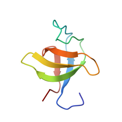
Second SH3 domain of ponsin solved from powder diffraction
Margiolaki, I., Wright, J.P., Wilmanns, M., Fitch, A.N., Pinotsis, N.(2007) J Am Chem Soc 129: 11865-11871
- PubMed: 17784760 Search on PubMed
- DOI: https://doi.org/10.1021/ja073846c
- Primary Citation Related Structures:
2O2W, 2O31 - PubMed Abstract:
Determination of protein crystal structures is dependent on the growth of high-quality single crystals, a process that is not always successful. Optimum crystallization conditions must be systematically sought for, and microcrystalline powders are frequently obtained in failed attempts to grow the desired crystal. In materials science, structures of samples ranging from ceramics, pharmaceuticals, zeolites, etc., can nowadays be solved, almost routinely, from powdered samples, and there seems to be no fundamental reason, except the sheer size and complexity of the structures involved, why powder diffraction should not be employed to solve structures of small proteins. Indeed, recent work has shown that the high-quality powder diffraction data can be used in the study of protein crystal structures. We report the solution, model building, and refinement of a 67-residue protein domain crystal structure, with a cell volume of 64 879 A3, from powder diffraction. The second SH3 domain of ponsin, a protein of high biological significance due to its role in cellular processes, is determined and refined to resolution limits comparable to single-crystal techniques. Our results demonstrate the power and future applicability of the powder technique in structural biology.
- European Synchrotron Radiation Facility, ESRF, BP-220, F-38043, Grenoble, France. margiolaki@esrf.fr
Organizational Affiliation: