2O2R

Crystal structure of the C-terminal domain of rat 10'formyltetrahydrofolate dehydrogenase in complex with NADPH


Experimental Data Snapshot

  • Method: X-RAY DIFFRACTION
  • Resolution: 2.20 Å
  • R-Value Free: 0.202 
  • R-Value Work: 0.180 
  • R-Value Observed: 0.181 

wwPDB Validation   3D Report Full Report


Ligand Structure Quality Assessment 


This is version 1.4 of the entry. See complete history


Literature

Crystal structures of the carboxyl terminal domain of rat 10-formyltetrahydrofolate dehydrogenase: implications for the catalytic mechanism of aldehyde dehydrogenases.

Tsybovsky, Y.Donato, H.Krupenko, N.I.Davies, C.Krupenko, S.A.

(2007) Biochemistry 46: 2917-2929

  • DOI: https://doi.org/10.1021/bi0619573
  • Primary Citation of Related Structures:  
    2O2P, 2O2Q, 2O2R

  • PubMed Abstract: 

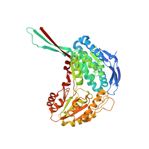
    10-Formyltetrahydrofolate dehydrogenase (FDH) catalyzes an NADP+-dependent dehydrogenase reaction resulting in conversion of 10-formyltetrahydrofolate to tetrahydrofolate and CO2. This reaction is a result of the concerted action of two catalytic domains of FDH, the amino-terminal hydrolase domain and the carboxyl-terminal aldehyde dehydrogenase domain. In addition to participation in the overall FDH mechanism, the C-terminal domain is capable of NADP+-dependent oxidation of short chain aldehydes to their corresponding acids. We have determined the crystal structure of the C-terminal domain of FDH and its complexes with oxidized and reduced forms of NADP. Compared to other members of the ALDH family, FDH demonstrates a new mode of binding of the 2'-phosphate group of NADP via a water-mediated contact with Gln600 that may contribute to the specificity of the enzyme for NADP over NAD. The structures also suggest how Glu673 can act as a general base in both acylation and deacylation steps of the reaction. In the apo structure, the general base Glu673 is positioned optimally for proton abstraction from the sulfur atom of Cys707. Upon binding of NADP+, the side chain of Glu673 is displaced from the active site by the nicotinamide ring and contacts a chain of highly ordered water molecules that may represent a pathway for translocation of the abstracted proton from Glu673 to the solvent. When reduced, the nicotinamide ring of NADP is displaced from the active site, restoring the contact between Cys707 and Glu673 and allowing the latter to activate the hydrolytic water molecule in deacylation.


  • Organizational Affiliation

    Department of Biochemistry and Molecular Biology, Medical University of South Carolina, Charleston, South Carolina 29425, USA.


Macromolecules
Find similar proteins by:  (by identity cutoff)  |  3D Structure
Entity ID: 1
MoleculeChains Sequence LengthOrganismDetailsImage
Formyltetrahydrofolate dehydrogenase
A, B, C, D
517Rattus norvegicusMutation(s): 0 
Gene Names: Fthfd
EC: 1.5.1.6
UniProt
Find proteins for P28037 (Rattus norvegicus)
Explore P28037 
Go to UniProtKB:  P28037
Entity Groups  
Sequence Clusters30% Identity50% Identity70% Identity90% Identity95% Identity100% Identity
UniProt GroupP28037
Sequence Annotations
Expand
  • Reference Sequence
Small Molecules
Ligands 3 Unique
IDChains Name / Formula / InChI Key2D Diagram3D Interactions
NDP
Query on NDP

Download Ideal Coordinates CCD File 
FA [auth D],
J [auth A],
Q [auth B],
Y [auth C]
NADPH DIHYDRO-NICOTINAMIDE-ADENINE-DINUCLEOTIDE PHOSPHATE
C21 H30 N7 O17 P3
ACFIXJIJDZMPPO-NNYOXOHSSA-N
SO4
Query on SO4

Download Ideal Coordinates CCD File 
AA [auth D]
BA [auth D]
CA [auth D]
DA [auth D]
E [auth A]
AA [auth D],
BA [auth D],
CA [auth D],
DA [auth D],
E [auth A],
EA [auth D],
F [auth A],
G [auth A],
H [auth A],
I [auth A],
L [auth B],
M [auth B],
N [auth B],
O [auth B],
P [auth B],
S [auth C],
T [auth C],
U [auth C],
V [auth C],
W [auth C],
X [auth C]
SULFATE ION
O4 S
QAOWNCQODCNURD-UHFFFAOYSA-L
GOL
Query on GOL

Download Ideal Coordinates CCD File 
GA [auth D],
K [auth A],
R [auth B],
Z [auth C]
GLYCEROL
C3 H8 O3
PEDCQBHIVMGVHV-UHFFFAOYSA-N
Experimental Data & Validation

Experimental Data

  • Method: X-RAY DIFFRACTION
  • Resolution: 2.20 Å
  • R-Value Free: 0.202 
  • R-Value Work: 0.180 
  • R-Value Observed: 0.181 
  • Space Group: C 1 2 1
Unit Cell:
Length ( Å )Angle ( ˚ )
a = 258.7α = 90
b = 194.3β = 108.9
c = 97.1γ = 90
Software Package:
Software NamePurpose
REFMACrefinement
MAR345data collection
HKL-2000data reduction
SCALEPACKdata scaling

Structure Validation

View Full Validation Report


Ligand Structure Quality Assessment 


Entry History 

Deposition Data

Revision History  (Full details and data files)

  • Version 1.0: 2007-03-06
    Type: Initial release
  • Version 1.1: 2008-05-01
    Changes: Version format compliance
  • Version 1.2: 2011-07-13
    Changes: Non-polymer description, Version format compliance
  • Version 1.3: 2017-10-18
    Changes: Refinement description
  • Version 1.4: 2023-08-30
    Changes: Data collection, Database references, Derived calculations, Refinement description