2O1V | pdb_00002o1v

Structure of full length GRP94 with ADP bound


Experimental Data Snapshot

  • Method: X-RAY DIFFRACTION
  • Resolution: 2.45 Å
  • R-Value Free: 
    0.299 (Depositor), 0.304 (DCC) 
  • R-Value Work: 
    0.248 (Depositor), 0.264 (DCC) 
  • R-Value Observed: 
    0.250 (Depositor) 

wwPDB Validation 3D Report Full Report

Validation slider image for 2O1V

Ligand Structure Quality Assessment 


This is version 1.4 of the entry. See complete history

Literature

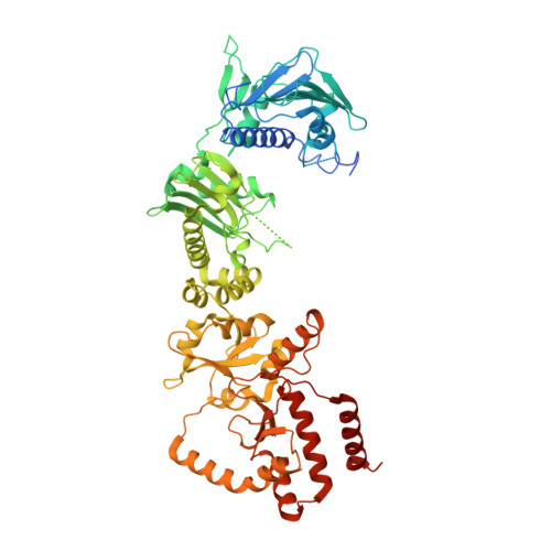
Structures of GRP94-Nucleotide Complexes Reveal Mechanistic Differences between the hsp90 Chaperones.

Dollins, D.E.Warren, J.J.Immormino, R.M.Gewirth, D.T.

(2007) Mol Cell 28: 41-56

  • DOI: https://doi.org/10.1016/j.molcel.2007.08.024
  • Primary Citation Related Structures: 
    2O1T, 2O1U, 2O1V, 2O1W

  • PubMed Abstract: 

    GRP94, an essential endoplasmic reticulum chaperone, is required for the conformational maturation of proteins destined for cell-surface display or export. The extent to which GRP94 and its cytosolic paralog, Hsp90, share a common mechanism remains controversial. GRP94 has not been shown conclusively to hydrolyze ATP or bind cochaperones, and both activities, by contrast, result in conformational changes and N-terminal dimerization in Hsp90 that are critical for its function. Here, we report the 2.4 A crystal structure of mammalian GRP94 in complex with AMPPNP and ADP. The chaperone is conformationally insensitive to the identity of the bound nucleotide, adopting a "twisted V" conformation that precludes N-terminal domain dimerization. We also present conclusive evidence that GRP94 possesses ATPase activity. Our observations provide a structural explanation for GRP94's observed rate of ATP hydrolysis and suggest a model for the role of ATP binding and hydrolysis in the GRP94 chaperone cycle.


  • Organizational Affiliation
    • Hauptman-Woodward Medical Research Institute, 700 Ellicott Street, Buffalo, NY 14203, USA.

Macromolecule Content 

  • Total Structure Weight: 154.14 kDa 
  • Atom Count: 9,607 
  • Modeled Residue Count: 1,153 
  • Deposited Residue Count: 1,332 
  • Unique protein chains: 1

Macromolecules

Find similar proteins by:|  3D Structure
Entity ID: 1
MoleculeChains  Sequence LengthOrganismDetailsImage
Endoplasmin
A, B
666Canis lupus familiarisMutation(s): 0 
Gene Names: HSP90B1TRA1
EC: 3.6.4
UniProt
Find proteins for P41148 (Canis lupus familiaris)
Explore P41148 
Go to UniProtKB:  P41148
Entity Groups
Sequence Clusters30% Identity50% Identity70% Identity90% Identity95% Identity100% Identity
UniProt GroupP41148
Sequence Annotations
Expand
Reference Sequence

Experimental Data & Validation

Experimental Data

  • Method: X-RAY DIFFRACTION
  • Resolution: 2.45 Å
  • R-Value Free:  0.299 (Depositor), 0.304 (DCC) 
  • R-Value Work:  0.248 (Depositor), 0.264 (DCC) 
  • R-Value Observed: 0.250 (Depositor) 
Space Group: P 21 21 21
Unit Cell:
Length ( Å )Angle ( ˚ )
a = 99.08α = 90
b = 108.85β = 90
c = 148.98γ = 90
Software Package:
Software NamePurpose
REFMACrefinement
ADSCdata collection
XDSdata reduction
XDSdata scaling
SHELXCDphasing
SHELXEmodel building

Structure Validation

View Full Validation Report


Ligand Structure Quality Assessment 


Entry History 

Deposition Data

Revision History  (Full details and data files)

  • Version 1.0: 2007-10-23
    Type: Initial release
  • Version 1.1: 2011-07-13
    Changes: Advisory, Derived calculations, Version format compliance
  • Version 1.2: 2017-08-02
    Changes: Source and taxonomy
  • Version 1.3: 2017-10-18
    Changes: Refinement description
  • Version 1.4: 2023-12-27
    Changes: Data collection, Database references, Derived calculations, Refinement description