2NU8 | pdb_00002nu8

C123aT Mutant of E. coli Succinyl-CoA Synthetase


Experimental Data Snapshot

  • Method: X-RAY DIFFRACTION
  • Resolution: 2.15 Å
  • R-Value Free: 
    0.255 (Depositor), 0.263 (DCC) 
  • R-Value Work: 
    0.210 (Depositor), 0.220 (DCC) 
  • R-Value Observed: 
    0.215 (Depositor) 

Starting Model: experimental
View more details

wwPDB Validation 3D Report Full Report

Validation slider image for 2NU8

Ligand Structure Quality Assessment 


This is version 1.5 of the entry. See complete history

Literature

Participation of Cys 123alpha of Escherichia coli Succinyl-CoA Synthetase in Catalysis

Hidber, E.Brownie, E.R.Hayakawa, K.Fraser, M.E.

(2007) Acta Crystallogr D Biol Crystallogr 63: 876-884

  • DOI: https://doi.org/10.1107/S0907444907029319
  • Primary Citation Related Structures: 
    2NU6, 2NU7, 2NU8, 2NU9, 2NUA

  • PubMed Abstract: 

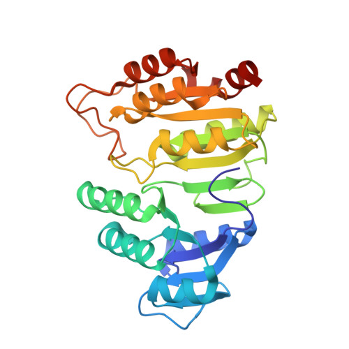
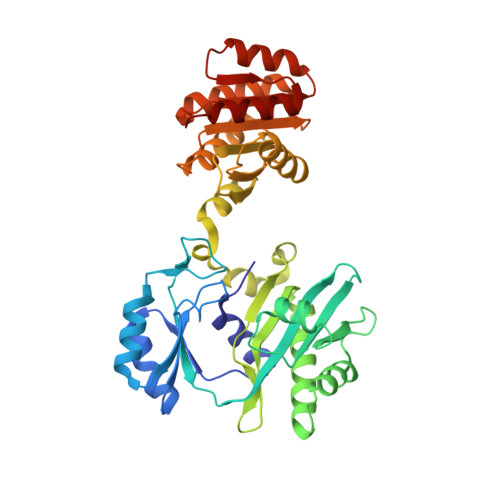
    Succinyl-CoA synthetase has a highly conserved cysteine residue, Cys123alpha in the Escherichia coli enzyme, that is located near the CoA-binding site and the active-site histidine residue. To test whether the succinyl moiety of succinyl-CoA is transferred to the thiol of Cys123alpha as part of the catalytic mechanism, this residue was mutated to alanine, serine, threonine and valine. Each mutant protein was catalytically active, although less active than the wild type. This proved that the specific formation of a thioester bond with Cys123alpha is not part of the catalytic mechanism. To understand why the mutations affected catalysis, the crystal structures of the four mutant proteins were determined. The alanine mutant showed no structural changes yet had reduced activity, suggesting that the size of the cysteine is important for optimal activity. These results explain why this cysteine residue is conserved in the sequences of succinyl-CoA synthetases from different sources.


  • Organizational Affiliation
    • Department of Biochemistry, University of Western Ontario, London, Ontario, Canada.

Macromolecule Content 

  • Total Structure Weight: 145.97 kDa 
  • Atom Count: 10,602 
  • Modeled Residue Count: 1,347 
  • Deposited Residue Count: 1,352 
  • Unique protein chains: 2

Macromolecules

Find similar proteins by:|  3D Structure
Entity ID: 1
MoleculeChains  Sequence LengthOrganismDetailsImage
Succinyl-CoA ligase [ADP-forming] subunit alphaA,
C [auth D]
288Escherichia coliMutation(s): 1 
Gene Names: sucD
EC: 6.2.1.5
UniProt
Find proteins for P0AGE9 (Escherichia coli (strain K12))
Explore P0AGE9 
Go to UniProtKB:  P0AGE9
Entity Groups
Sequence Clusters30% Identity50% Identity70% Identity90% Identity95% Identity100% Identity
UniProt GroupP0AGE9
Sequence Annotations
Expand
Reference Sequence
Find similar proteins by:|  3D Structure
Entity ID: 2
MoleculeChains  Sequence LengthOrganismDetailsImage
Succinyl-CoA synthetase beta chainB,
D [auth E]
388Escherichia coliMutation(s): 0 
Gene Names: sucC
EC: 6.2.1.5
UniProt
Find proteins for P0A836 (Escherichia coli (strain K12))
Explore P0A836 
Go to UniProtKB:  P0A836
Entity Groups
Sequence Clusters30% Identity50% Identity70% Identity90% Identity95% Identity100% Identity
UniProt GroupP0A836
Sequence Annotations
Expand
Reference Sequence

Small Molecules

Ligands 4 Unique
IDChains Name / Formula / InChI Key2D Diagram3D Interactions
COA

Query on COA


Download:Ideal Coordinates CCD File
G [auth A],
J [auth B],
L [auth D],
O [auth E]
COENZYME A
C21 H36 N7 O16 P3 S
RGJOEKWQDUBAIZ-IBOSZNHHSA-N
SO4

Query on SO4


Download:Ideal Coordinates CCD File
F [auth A],
I [auth B],
M [auth E],
N [auth E]
SULFATE ION
O4 S
QAOWNCQODCNURD-UHFFFAOYSA-L
PO4

Query on PO4


Download:Ideal Coordinates CCD File
E [auth A],
K [auth D]
PHOSPHATE ION
O4 P
NBIIXXVUZAFLBC-UHFFFAOYSA-K
GOL

Query on GOL


Download:Ideal Coordinates CCD File
H [auth A]GLYCEROL
C3 H8 O3
PEDCQBHIVMGVHV-UHFFFAOYSA-N

Experimental Data & Validation

Experimental Data

  • Method: X-RAY DIFFRACTION
  • Resolution: 2.15 Å
  • R-Value Free:  0.255 (Depositor), 0.263 (DCC) 
  • R-Value Work:  0.210 (Depositor), 0.220 (DCC) 
  • R-Value Observed: 0.215 (Depositor) 
Space Group: P 43 2 2
Unit Cell:
Length ( Å )Angle ( ˚ )
a = 96.69α = 90
b = 96.69β = 90
c = 385.94γ = 90
Software Package:
Software NamePurpose
ADSCdata collection
CNSrefinement
DENZOdata reduction
SCALEPACKdata scaling

Structure Validation

View Full Validation Report


Ligand Structure Quality Assessment 


Entry History 

Deposition Data

  • Released Date: 2007-07-24 
  • Deposition Author(s): Fraser, M.E.

Revision History  (Full details and data files)

  • Version 1.0: 2007-07-24
    Type: Initial release
  • Version 1.1: 2008-05-01
    Changes: Version format compliance
  • Version 1.2: 2011-07-13
    Changes: Non-polymer description, Version format compliance
  • Version 1.3: 2021-10-20
    Changes: Database references, Derived calculations
  • Version 1.4: 2023-08-30
    Changes: Data collection, Refinement description
  • Version 1.5: 2024-10-30
    Changes: Structure summary