2NST

Crystal structure of pectin methylesterase D178A mutant in complex with hexasaccharide II


Experimental Data Snapshot

  • Method: X-RAY DIFFRACTION
  • Resolution: 1.70 Å
  • R-Value Free: 0.231 
  • R-Value Work: 0.200 
  • R-Value Observed: 0.201 

wwPDB Validation   3D Report Full Report


This is version 3.2 of the entry. See complete history


Literature

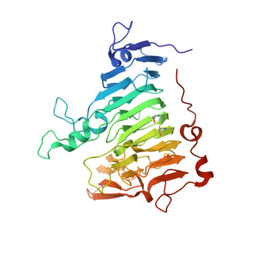
Molecular basis of the activity of the phytopathogen pectin methylesterase.

Fries, M.Ihrig, J.Brocklehurst, K.Shevchik, V.E.Pickersgill, R.W.

(2007) EMBO J 26: 3879-3887

  • DOI: https://doi.org/10.1038/sj.emboj.7601816
  • Primary Citation of Related Structures:  
    2NSP, 2NST, 2NT6, 2NT9, 2NTB, 2NTP, 2NTQ

  • PubMed Abstract: 

    We provide a mechanism for the activity of pectin methylesterase (PME), the enzyme that catalyses the essential first step in bacterial invasion of plant tissues. The complexes formed in the crystal using specifically methylated pectins, together with kinetic measurements of directed mutants, provide clear insights at atomic resolution into the specificity and the processive action of the Erwinia chrysanthemi enzyme. Product complexes provide additional snapshots along the reaction coordinate. We previously revealed that PME is a novel aspartic-esterase possessing parallel beta-helix architecture and now show that the two conserved aspartates are the nucleophile and general acid-base in the mechanism, respectively. Other conserved residues at the catalytic centre are shown to be essential for substrate binding or transition state stabilisation. The preferential binding of methylated sugar residues upstream of the catalytic site, and demethylated residues downstream, drives the enzyme along the pectin molecule and accounts for the sequential pattern of demethylation produced by both bacterial and plant PMEs.


  • Organizational Affiliation

    School of Biological and Chemical Sciences, Queen Mary, University of London, London, UK.


Macromolecules
Find similar proteins by:  (by identity cutoff)  |  3D Structure
Entity ID: 1
MoleculeChains Sequence LengthOrganismDetailsImage
Pectinesterase A
A, B
342Dickeya dadantii 3937Mutation(s): 1 
Gene Names: pemApem
EC: 3.1.1.11
UniProt
Find proteins for P0C1A9 (Dickeya dadantii (strain 3937))
Explore P0C1A9 
Go to UniProtKB:  P0C1A9
Entity Groups  
Sequence Clusters30% Identity50% Identity70% Identity90% Identity95% Identity100% Identity
UniProt GroupP0C1A9
Sequence Annotations
Expand
  • Reference Sequence
Oligosaccharides

Help

Entity ID: 2
MoleculeChains Length2D Diagram Glycosylation3D Interactions
alpha-D-galactopyranuronic acid-(1-4)-alpha-D-galactopyranuronic acid-(1-4)-methyl alpha-D-galactopyranuronate-(1-4)-methyl alpha-D-galactopyranuronate-(1-4)-methyl alpha-D-galactopyranuronate-(1-4)-alpha-D-galactopyranuronic acid
C, D
6N/A
Glycosylation Resources
GlyTouCan:  G41087WO
GlyCosmos:  G41087WO
Experimental Data & Validation

Experimental Data

  • Method: X-RAY DIFFRACTION
  • Resolution: 1.70 Å
  • R-Value Free: 0.231 
  • R-Value Work: 0.200 
  • R-Value Observed: 0.201 
  • Space Group: P 1 21 1
Unit Cell:
Length ( Å )Angle ( ˚ )
a = 51.012α = 90
b = 84.84β = 93.57
c = 97.163γ = 90
Software Package:
Software NamePurpose
REFMACrefinement
MOSFLMdata reduction
SCALAdata scaling
REFMACphasing

Structure Validation

View Full Validation Report


Entry History 

Deposition Data

Revision History  (Full details and data files)

  • Version 1.0: 2007-09-18
    Type: Initial release
  • Version 1.1: 2011-07-13
    Changes: Source and taxonomy, Version format compliance
  • Version 2.0: 2020-05-13
    Changes: Atomic model, Data collection, Derived calculations
  • Version 3.0: 2020-07-29
    Type: Remediation
    Reason: Carbohydrate remediation
    Changes: Atomic model, Data collection, Derived calculations, Structure summary
  • Version 3.1: 2021-10-20
    Changes: Database references, Derived calculations, Structure summary
  • Version 3.2: 2023-08-30
    Changes: Data collection, Refinement description