2NS6 | pdb_00002ns6

Crystal Structure of the Minimal Relaxase Domain of MobA from Plasmid R1162


Experimental Data Snapshot

  • Method: X-RAY DIFFRACTION
  • Resolution: 2.10 Å
  • R-Value Free: 
    0.287 (Depositor), 0.222 (DCC) 
  • R-Value Work: 
    0.238 (Depositor), 0.240 (DCC) 

wwPDB Validation 3D Report Full Report

Validation slider image for 2NS6

This is version 1.4 of the entry. See complete history

Literature

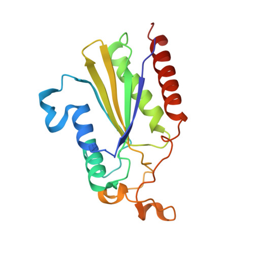
The Structure of the Minimal Relaxase Domain of MobA at 2.1 A Resolution.

Monzingo, A.F.Ozburn, A.Xia, S.Meyer, R.J.Robertus, J.D.

(2007) J Mol Biology 366: 165-178

  • DOI: https://doi.org/10.1016/j.jmb.2006.11.031
  • Primary Citation Related Structures: 
    2NS6

  • PubMed Abstract: 

    The plasmid R1162 encodes proteins that enable its conjugative mobilization between bacterial cells. It can transfer between many different species and is one of the most promiscuous of the mobilizable plasmids. The plasmid-encoded protein MobA, which has both nicking and priming activities on single-stranded DNA, is essential for mobilization. The nicking, or relaxase, activity has been localized to the 186 residue N-terminal domain, called minMobA. We present here the 2.1 A X-ray structure of minMobA. The fold is similar to that seen for two other relaxases, TraI and TrwC. The similarity in fold, and action, suggests these enzymes are evolutionary homologs, despite the lack of any significant amino acid similarity. MinMobA has a well- defined target DNA called oriT. The active site metal is observed near Tyr25, which is known to form a phosphotyrosine adduct with the substrate. A model of the oriT substrate complexed with minMobA has been made, based on observed substrate binding to TrwC and TraI. The model is consistent with observations of substrate base specificity, and provides a rationalization for elements of the likely enzyme mechanism.


  • Organizational Affiliation
    • Institute of Cellular and Molecular Biology, Department of Chemistry and Biochemistry, 1 University Station A5300, University of Texas, Austin, TX 78712, USA.

Macromolecule Content 

  • Total Structure Weight: 20.98 kDa 
  • Atom Count: 1,556 
  • Modeled Residue Count: 183 
  • Deposited Residue Count: 185 
  • Unique protein chains: 1

Macromolecules

Find similar proteins by:|  3D Structure
Entity ID: 1
MoleculeChains  Sequence LengthOrganismDetailsImage
Mobilization protein A185Pseudomonas aeruginosaMutation(s): 0 
Gene Names: mobA
EC: 3.6.1
UniProt
Find proteins for D0VWX2 (Pseudomonas aeruginosa)
Explore D0VWX2 
Go to UniProtKB:  D0VWX2
Entity Groups
Sequence Clusters30% Identity50% Identity70% Identity90% Identity95% Identity100% Identity
UniProt GroupD0VWX2
Sequence Annotations
Expand
Reference Sequence

Experimental Data & Validation

Experimental Data

  • Method: X-RAY DIFFRACTION
  • Resolution: 2.10 Å
  • R-Value Free:  0.287 (Depositor), 0.222 (DCC) 
  • R-Value Work:  0.238 (Depositor), 0.240 (DCC) 
Space Group: P 41 21 2
Unit Cell:
Length ( Å )Angle ( ˚ )
a = 47.967α = 90
b = 47.967β = 90
c = 165.927γ = 90
Software Package:
Software NamePurpose
DENZOdata reduction
SCALEPACKdata scaling
CNSrefinement
PDB_EXTRACTdata extraction
HKL-2000data reduction
SOLVEphasing

Structure Validation

View Full Validation Report


Entry History 

Deposition Data

Revision History  (Full details and data files)

  • Version 1.0: 2007-02-06
    Type: Initial release
  • Version 1.1: 2008-01-10
    Changes: Version format compliance
  • Version 1.2: 2011-07-13
    Changes: Version format compliance
  • Version 1.3: 2017-10-18
    Changes: Refinement description
  • Version 1.4: 2023-12-27
    Changes: Data collection, Database references, Derived calculations