2NOT | pdb_00002not

NOTECHIS II-5, NEUROTOXIC PHOSPHOLIPASE A2 FROM NOTECHIS SCUTATUS SCUTATUS


Experimental Data Snapshot

  • Method: X-RAY DIFFRACTION
  • Resolution: 3.00 Å
  • R-Value Free: 
    0.255 (Depositor) 
  • R-Value Work: 
    0.219 (Depositor), 0.204 (DCC) 
  • R-Value Observed: 
    0.219 (Depositor) 

Starting Model: experimental
View more details

wwPDB Validation 3D Report Full Report

Validation slider image for 2NOT

This is version 1.4 of the entry. See complete history

Literature

The three-dimensional structures of two toxins from snake venom throw light on the anticoagulant and neurotoxic sites of phospholipase A2.

Carredano, E.Westerlund, B.Persson, B.Saarinen, M.Ramaswamy, S.Eaker, D.Eklund, H.

(1998) Toxicon 36: 75-92

  • DOI: https://doi.org/10.1016/s0041-0101(97)00051-2
  • Primary Citation Related Structures: 
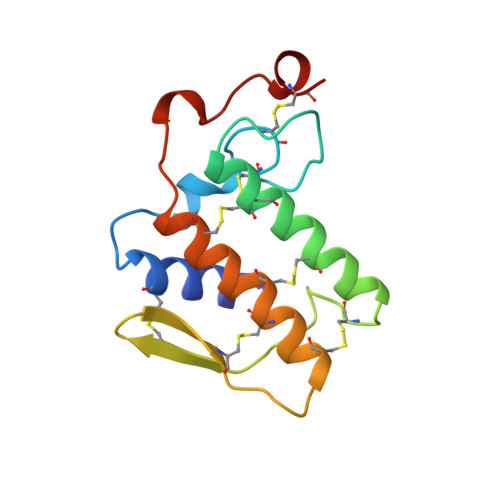
    1VIP, 2NOT

  • PubMed Abstract: 

    The three-dimensional structures of the class II anticoagulant phospholipase A2 (PLA2) toxin RVV-VD from the venom of Russell's viper, Vipera russelli russelli, and the class I neurotoxic PLA2 Notechis II-5 from the, Australian tiger snake, Notechis scutatus scutatus, were determined to 2.2 A and 3.0 A resolution, respectively. Both enzymes are monomeric and consist of 121 and 119 residues, respectively. A comparison of ten class I/II PLA2 structures showed, among other differences, that the beta-sheet of these enzymes (residues 76-83) is about 90 degrees less twisted in class I than in class II PLA2s. This, along with the insertion of some residues in the region 57-59 in class I enzymes (the elapid loop), could be the main reason for the significant difference in the anticoagulant and (presynaptic) neurotoxic properties between the two classes of PLA2. It seems apparent from sequence and structural comparisons that the toxic site of PLA2 responsible for the strong anticoagulancy of these toxins consists of a negatively charged part, Glu53, together with a positively charged ridge of lysine residues free for intermolecular interactions. These lysines differ between the two classes of PLA2.


  • Organizational Affiliation
    • Department of Molecular Biology, Swedish University of Agricultural Sciences, Uppsala, Sweden.

Macromolecule Content 

  • Total Structure Weight: 27.39 kDa 
  • Atom Count: 1,912 
  • Modeled Residue Count: 238 
  • Deposited Residue Count: 238 
  • Unique protein chains: 1

Macromolecules

Find similar proteins by:|  3D Structure
Entity ID: 1
MoleculeChains  Sequence LengthOrganismDetailsImage
PHOSPHOLIPASE A2
A, B
119Notechis scutatus scutatusMutation(s): 0 
EC: 3.1.1.4
UniProt
Find proteins for P00609 (Notechis scutatus scutatus)
Explore P00609 
Go to UniProtKB:  P00609
Entity Groups
Sequence Clusters30% Identity50% Identity70% Identity90% Identity95% Identity100% Identity
UniProt GroupP00609
Sequence Annotations
Expand
Reference Sequence

Experimental Data & Validation

Experimental Data

  • Method: X-RAY DIFFRACTION
  • Resolution: 3.00 Å
  • R-Value Free:  0.255 (Depositor) 
  • R-Value Work:  0.219 (Depositor), 0.204 (DCC) 
  • R-Value Observed: 0.219 (Depositor) 
Space Group: P 21 21 21
Unit Cell:
Length ( Å )Angle ( ˚ )
a = 39.5α = 90
b = 43.5β = 90
c = 145γ = 90
Software Package:
Software NamePurpose
AMoREphasing
X-PLORrefinement

Structure Validation

View Full Validation Report


Entry History 

Deposition Data

Revision History  (Full details and data files)

  • Version 1.0: 1997-06-16
    Type: Initial release
  • Version 1.1: 2008-03-03
    Changes: Version format compliance
  • Version 1.2: 2011-07-13
    Changes: Version format compliance
  • Version 1.3: 2018-04-04
    Changes: Data collection
  • Version 1.4: 2024-10-30
    Changes: Data collection, Database references, Refinement description, Structure summary