2NLX | pdb_00002nlx

Crystal structure of the apo E. coli xylulose kinase


Experimental Data Snapshot

  • Method: X-RAY DIFFRACTION
  • Resolution: 2.70 Å
  • R-Value Free: 
    0.254 (Depositor), 0.247 (DCC) 
  • R-Value Work: 
    0.205 (Depositor), 0.201 (DCC) 
  • R-Value Observed: 
    0.207 (Depositor) 

wwPDB Validation 3D Report Full Report

Validation slider image for 2NLX

This is version 1.5 of the entry. See complete history

Literature

Structural and kinetic studies of induced fit in xylulose kinase from Escherichia coli.

Di Luccio, E.Petschacher, B.Voegtli, J.Chou, H.T.Stahlberg, H.Nidetzky, B.Wilson, D.K.

(2007) J Mol Biology 365: 783-798

  • DOI: https://doi.org/10.1016/j.jmb.2006.10.068
  • Primary Citation Related Structures: 
    2ITM, 2NLX

  • PubMed Abstract: 

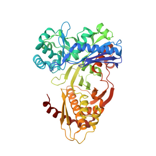
    The primary metabolic route for D-xylose, the second most abundant sugar in nature, is via the pentose phosphate pathway after a two-step or three-step conversion to xylulose-5-phosphate. Xylulose kinase (XK; EC 2.7.1.17) phosphorylates D-xylulose, the last step in this conversion. The apo and D-xylulose-bound crystal structures of Escherichia coli XK have been determined and show a dimer composed of two domains separated by an open cleft. XK dimerization was observed directly by a cryo-EM reconstruction at 36 A resolution. Kinetic studies reveal that XK has a weak substrate-independent MgATP-hydrolyzing activity, and phosphorylates several sugars and polyols with low catalytic efficiency. Binding of pentulose and MgATP to form the reactive ternary complex is strongly synergistic. Although the steady-state kinetic mechanism of XK is formally random, a path is preferred in which D-xylulose binds before MgATP. Modelling of MgATP binding to XK and the accompanying conformational change suggests that sugar binding is accompanied by a dramatic hinge-bending movement that enhances interactions with MgATP, explaining the observed synergism. A catalytic mechanism is proposed and supported by relevant site-directed mutants.


  • Organizational Affiliation
    • Section of Molecular and Cellular Biology, University of California, Davis, CA 95616, USA.

Macromolecule Content 

  • Total Structure Weight: 105.34 kDa 
  • Atom Count: 7,394 
  • Modeled Residue Count: 948 
  • Deposited Residue Count: 968 
  • Unique protein chains: 1

Macromolecules

Find similar proteins by:|  3D Structure
Entity ID: 1
MoleculeChains  Sequence LengthOrganismDetailsImage
Xylulose kinase
A, B
484Escherichia coliMutation(s): 0 
Gene Names: xylB
EC: 2.7.1.17 (PDB Primary Data), 2.7.1 (UniProt)
UniProt
Find proteins for P09099 (Escherichia coli (strain K12))
Explore P09099 
Go to UniProtKB:  P09099
Entity Groups
Sequence Clusters30% Identity50% Identity70% Identity90% Identity95% Identity100% Identity
UniProt GroupP09099
Sequence Annotations
Expand
Reference Sequence

Experimental Data & Validation

Experimental Data

  • Method: X-RAY DIFFRACTION
  • Resolution: 2.70 Å
  • R-Value Free:  0.254 (Depositor), 0.247 (DCC) 
  • R-Value Work:  0.205 (Depositor), 0.201 (DCC) 
  • R-Value Observed: 0.207 (Depositor) 
Space Group: P 21 21 21
Unit Cell:
Length ( Å )Angle ( ˚ )
a = 67.78α = 90
b = 112.02β = 90
c = 138.8γ = 90
Software Package:
Software NamePurpose
DENZOdata reduction
SCALEPACKdata scaling
REFMACrefinement
PDB_EXTRACTdata extraction
CrystalCleardata collection
HKL-2000data reduction
CNSphasing

Structure Validation

View Full Validation Report


Entry History 

Deposition Data

Revision History  (Full details and data files)

  • Version 1.0: 2006-11-14
    Type: Initial release
  • Version 1.1: 2008-05-01
    Changes: Version format compliance
  • Version 1.2: 2011-07-13
    Changes: Version format compliance
  • Version 1.3: 2017-10-18
    Changes: Refinement description
  • Version 1.4: 2018-01-31
    Changes: Experimental preparation
  • Version 1.5: 2023-12-27
    Changes: Data collection, Database references, Refinement description