Determination of ligand binding modes in weak protein-ligand complexes using sparse NMR data.
Mohanty, B., Williams, M.L., Doak, B.C., Vazirani, M., Ilyichova, O., Wang, G., Bermel, W., Simpson, J.S., Chalmers, D.K., King, G.F., Mobli, M., Scanlon, M.J.(2016) J Biomol NMR 66: 195-208
- PubMed: 27778134 Search on PubMed
- DOI: https://doi.org/10.1007/s10858-016-0067-4
- Primary Citation Related Structures:
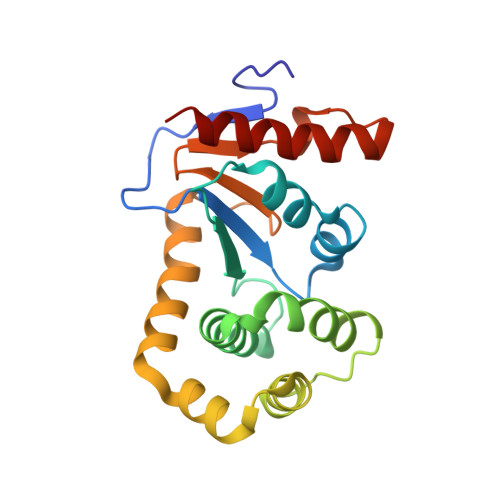
2NDO, 4ZIJ - PubMed Abstract:
We describe a general approach to determine the binding pose of small molecules in weakly bound protein-ligand complexes by deriving distance constraints between the ligand and methyl groups from all methyl-containing residues of the protein. We demonstrate that using a single sample, which can be prepared without the use of expensive precursors, it is possible to generate high-resolution data rapidly and obtain the resonance assignments of Ile, Leu, Val, Ala and Thr methyl groups using triple resonance scalar correlation data. The same sample may be used to obtain Met ε CH 3 assignments using NOESY-based methods, although the superior sensitivity of NOESY using [U- 13 C, 15 N]-labeled protein makes the use of this second sample more efficient. We describe a structural model for a weakly binding ligand bound to its target protein, DsbA, derived from intermolecular methyl-to-ligand nuclear Overhauser enhancements, and demonstrate that the ability to assign all methyl resonances in the spectrum is essential to derive an accurate model of the structure. Once the methyl assignments have been obtained, this approach provides a rapid means to generate structural models for weakly bound protein-ligand complexes. Such weak complexes are often found at the beginning of programs of fragment based drug design and can be challenging to characterize using X-ray crystallography.
- Medicinal Chemistry, Monash Institute of Pharmaceutical Sciences, Monash University, 381 Royal Parade, Parkville, VIC, 3052, Australia.
Organizational Affiliation: