2N77

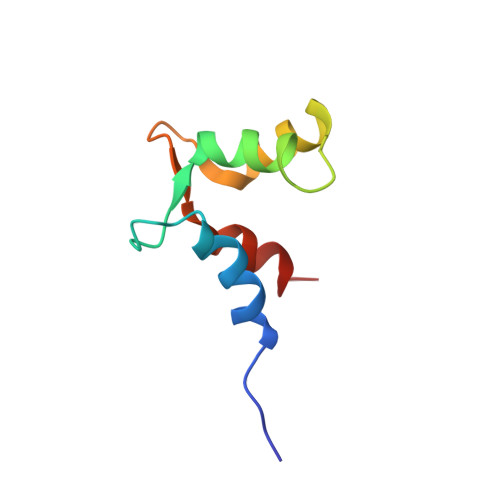
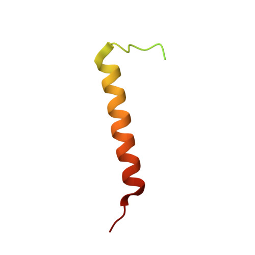
NMR solution structure of a complex of PEP-19 bound to the C-domain of apo calmodulin


Experimental Data Snapshot

  • Method: SOLUTION NMR
  • Conformers Calculated: 300 
  • Conformers Submitted: 20 
  • Selection Criteria: structures with the lowest energy 

wwPDB Validation   3D Report Full Report


This is version 1.3 of the entry. See complete history


Literature

PEP-19 modulates calcium binding to calmodulin by electrostatic steering.

Wang, X.Putkey, J.A.

(2016) Nat Commun 7: 13583-13583

  • DOI: https://doi.org/10.1038/ncomms13583
  • Primary Citation of Related Structures:  
    2N77

  • PubMed Abstract: 

    PEP-19 is a small protein that increases the rates of Ca 2+ binding to the C-domain of calmodulin (CaM) by an unknown mechanism. Although an IQ motif promotes binding to CaM, an acidic sequence in PEP-19 is required to modulate Ca 2+ binding and to sensitize HeLa cells to ATP-induced Ca 2+ release. Here, we report the NMR solution structure of a complex between PEP-19 and the C-domain of apo CaM. The acidic sequence of PEP-19 associates between helices E and F of CaM via hydrophobic interactions. This allows the acidic side chains in PEP-19 to extend toward the solvent and form a negatively charged surface that resembles a catcher's mitt near Ca 2+ binding loop III of CaM. The topology and gradients of negative electrostatic surface potential support a mechanism by which PEP-19 increases the rate of Ca 2+ binding to the C-domain of CaM by 'catching' and electrostatically steering Ca 2+ to site III.


  • Organizational Affiliation

    Department of Biochemistry and Molecular Biology, McGovern Medical at UTHealth, 6431 Fannin, Houston, Texas 77030, USA.


Macromolecules
Find similar proteins by:  (by identity cutoff)  |  3D Structure
Entity ID: 1
MoleculeChains Sequence LengthOrganismDetailsImage
Calmodulin73Homo sapiensMutation(s): 0 
Gene Names: 
CALM1CALMCAMCAM1CALM2CAM2CAMBCALM3CALML2CAM3...
CALM1CALMCAMCAM1CALM2CAM2CAMBCALM3CALML2CAM3CAMCCAMIII

UniProt & NIH Common Fund Data Resources
Find proteins for P0DP23 (Homo sapiens)
Explore P0DP23 
Go to UniProtKB:  P0DP23
PHAROS:  P0DP23
GTEx:  ENSG00000198668 
Entity Groups  
Sequence Clusters30% Identity50% Identity70% Identity90% Identity95% Identity100% Identity
UniProt GroupP0DP23
Sequence Annotations
Expand
  • Reference Sequence
Find similar proteins by:  (by identity cutoff)  |  3D Structure
Entity ID: 2
MoleculeChains Sequence LengthOrganismDetailsImage
Purkinje cell protein 462Homo sapiensMutation(s): 0 
Gene Names: PCP4PEP19
UniProt & NIH Common Fund Data Resources
Find proteins for P48539 (Homo sapiens)
Explore P48539 
Go to UniProtKB:  P48539
PHAROS:  P48539
GTEx:  ENSG00000183036 
Entity Groups  
Sequence Clusters30% Identity50% Identity70% Identity90% Identity95% Identity100% Identity
UniProt GroupP48539
Sequence Annotations
Expand
  • Reference Sequence
Experimental Data & Validation

Experimental Data

  • Method: SOLUTION NMR
  • Conformers Calculated: 300 
  • Conformers Submitted: 20 
  • Selection Criteria: structures with the lowest energy 

Structure Validation

View Full Validation Report


Entry History 

Deposition Data

Revision History  (Full details and data files)

  • Version 1.0: 2016-11-30
    Type: Initial release
  • Version 1.1: 2016-12-07
    Changes: Database references
  • Version 1.2: 2021-08-18
    Changes: Data collection, Database references, Experimental preparation
  • Version 1.3: 2024-05-15
    Changes: Data collection, Database references