2MZP

Structure and dynamics of the acidosis-resistant a162H mutant of the switch region of troponin I bound to the regulatory domain of troponin C


Experimental Data Snapshot

  • Method: SOLUTION NMR
  • Conformers Calculated: 200 
  • Conformers Submitted: 20 
  • Selection Criteria: structures with the lowest energy 

wwPDB Validation   3D Report Full Report


This is version 1.2 of the entry. See complete history


Literature

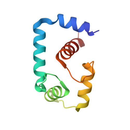
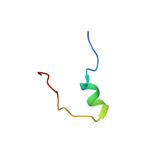
Structure and Dynamics of the Acidosis-Resistant A162H Mutant of the Switch Region of Troponin I Bound to the Regulatory Domain of Troponin C.

Pineda-Sanabria, S.E.Robertson, I.M.Sykes, B.D.

(2015) Biochemistry 54: 3583-3593

  • DOI: https://doi.org/10.1021/acs.biochem.5b00178
  • Primary Citation of Related Structures:  
    2MZP

  • PubMed Abstract: 

    Intracellular acidosis lowers the Ca²⁺ sensitivity of cardiac muscle, which results in decreased force generation, decreased cardiac output, and, eventually, heart failure. The A162H mutant of cardiac troponin I in the thin filament turns the heart acidosis-resistant. Physiological and structural studies have provided insights into the mechanism of protection by the A162H substitution; however, the effect of other native residues of cardiac troponin I is not fully understood. In this study, we determined the structure of the A162H mutant of the switch region of cardiac troponin I bound to the regulatory domain of troponin C at pH 6.1, and the dynamics as a function of pH, by NMR spectroscopy to evaluate the changes induced by protonation of A162H. The results indicate that A162H induces a transitory curved conformation on troponin I that promotes contraction, but it is countered by residue E164 to ensure proper relaxation. Our model explains the absence of diastolic impairment in the gain-of-function phenotype induced by the A162H substitution as well as the effects of a variety of mutants studied previously. The description of this mechanism underlines the fine quality of regulation on cardiac muscle contraction and anticipates pharmacological agents that induce modest changes in the contraction-relaxation equilibrium to produce marked effects in cardiac performance.


Macromolecules
Find similar proteins by:  (by identity cutoff)  |  3D Structure
Entity ID: 1
MoleculeChains Sequence LengthOrganismDetailsImage
Troponin C, slow skeletal and cardiac musclesA [auth C]89Homo sapiensMutation(s): 0 
Gene Names: TNNC1TNNC
UniProt & NIH Common Fund Data Resources
Find proteins for P63316 (Homo sapiens)
Explore P63316 
Go to UniProtKB:  P63316
PHAROS:  P63316
GTEx:  ENSG00000114854 
Entity Groups  
Sequence Clusters30% Identity50% Identity70% Identity90% Identity95% Identity100% Identity
UniProt GroupP63316
Sequence Annotations
Expand
  • Reference Sequence
Find similar proteins by:  (by identity cutoff)  |  3D Structure
Entity ID: 2
MoleculeChains Sequence LengthOrganismDetailsImage
Troponin I, cardiac muscleB [auth I]27Homo sapiensMutation(s): 1 
UniProt & NIH Common Fund Data Resources
Find proteins for P19429 (Homo sapiens)
Explore P19429 
Go to UniProtKB:  P19429
PHAROS:  P19429
GTEx:  ENSG00000129991 
Entity Groups  
Sequence Clusters30% Identity50% Identity70% Identity90% Identity95% Identity100% Identity
UniProt GroupP19429
Sequence Annotations
Expand
  • Reference Sequence
Small Molecules
Ligands 1 Unique
IDChains Name / Formula / InChI Key2D Diagram3D Interactions
CA
Query on CA

Download Ideal Coordinates CCD File 
C
CALCIUM ION
Ca
BHPQYMZQTOCNFJ-UHFFFAOYSA-N
Experimental Data & Validation

Experimental Data

  • Method: SOLUTION NMR
  • Conformers Calculated: 200 
  • Conformers Submitted: 20 
  • Selection Criteria: structures with the lowest energy 

Structure Validation

View Full Validation Report


Entry History 

Deposition Data

Revision History  (Full details and data files)

  • Version 1.0: 2015-06-03
    Type: Initial release
  • Version 1.1: 2015-07-01
    Changes: Database references
  • Version 1.2: 2023-06-14
    Changes: Data collection, Database references, Derived calculations, Other