Nuclear Magnetic Resonance Structure of the III-IV-V Three-Way Junction from the Varkud Satellite Ribozyme and Identification of Magnesium-Binding Sites Using Paramagnetic Relaxation Enhancement.
Bonneau, E., Legault, P.(2014) Biochemistry 53: 6264-6275
- PubMed: 25238589 Search on PubMed
- DOI: https://doi.org/10.1021/bi500826n
- Primary Citation Related Structures:
2MTJ, 2MTK - PubMed Abstract:
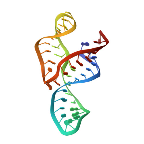
The VS ribozyme is a catalytic RNA found within some natural isolates of Neurospora that is being used as a model system to improve our understanding of RNA structure, catalysis, and engineering. The catalytic domain contains five helical domains (SLII-SLVI) that are organized by two three-way junctions. The III-IV-V junction is required for high-affinity binding of the substrate domain (SLI) through formation of a kissing loop interaction with SLV. Here, we determine the high-resolution nuclear magnetic resonance (NMR) structure of a 47-nucleotide RNA containing the III-IV-V junction (J345). The J345 RNA adopts a Y-shaped fold typical of the family C three-way junctions, with coaxial stacking between stems III and IV and an acute angle between stems III and V. The NMR structure reveals that the core of the III-IV-V junction contains four stacked base triples, a U-turn motif, a cross-strand stacking interaction, an A-minor interaction, and a ribose zipper. In addition, the NMR structure shows that the cCUUGg tetraloop used to stabilize stem IV adopts a novel RNA tetraloop fold, different from the known gCUUGc tetraloop structure. Using Mn(2+)-induced paramagnetic relaxation enhancement, we identify six Mg(2+)-binding sites within J345, including one associated with the cCUUGg tetraloop and two with the junction core. The NMR structure of J345 likely represents the conformation of the III-IV-V junction in the context of the active VS ribozyme and suggests that this junction functions as a dynamic hinge that contributes to substrate recognition and catalysis. Moreover, this study highlights a new role for family C three-way junctions in long-range tertiary interactions.
- Département de Biochimie et Médecine Moléculaire, Université de Montréal , C.P. 6128, Succursale Centre-Ville, Montréal, QC, Canada H3C 3J7.
Organizational Affiliation: