2MB0 | pdb_00002mb0

Solution structure of hnRNP G RRM in complex with the RNA 5'-AUCAAA-3'


Experimental Data Snapshot

  • Method: SOLUTION NMR
  • Conformers Calculated: 50 
  • Conformers Submitted: 20 
  • Selection Criteria: structures with the lowest energy 

wwPDB Validation 3D Report Full Report

Validation slider image for 2MB0

This is version 1.3 of the entry. See complete history

Literature

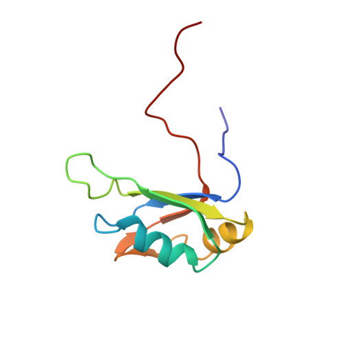
Characterization of the RNA recognition mode of hnRNP G extends its role in SMN2 splicing regulation.

Moursy, A.Allain, F.H.Clery, A.

(2014) Nucleic Acids Res 42: 6659-6672

  • DOI: https://doi.org/10.1093/nar/gku244
  • Primary Citation Related Structures: 
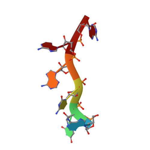
    2MB0

  • PubMed Abstract: 

    Regulation of SMN2 exon 7 splicing is crucial for the production of active SMN protein and the survival of Spinal Muscular Atrophy (SMA) patients. One of the most efficient activators of exon 7 inclusion is hnRNP G, which is recruited to the exon by Tra2-β1. We report that in addition to the C-terminal region of hnRNP G, the RNA Recognition Motif (RRM) and the middle part of the protein containing the Arg-Gly-Gly (RGG) box are important for this function. To better understand the mode of action of hnRNP G in this context we determined the structure of its RRM bound to an SMN2 derived RNA. The RRM interacts with a 5'-AAN-3' motif and specifically recognizes the two consecutive adenines. By testing the effect of mutations in hnRNP G RRM and in its putative binding sites on the splicing of SMN2 exon 7, we show that it specifically binds to exon 7. This interaction is required for hnRNP G splicing activity and we propose its recruitment to a polyA tract located upstream of the Tra2-β1 binding site. Finally, our data suggest that hnRNP G plays a major role in the recruitment of the Tra2-β1/hnRNP G/SRSF9 trimeric complex to SMN2 exon 7.


  • Organizational Affiliation
    • Institute for Molecular Biology and Biophysics, Swiss Federal Institute of Technology (ETH), 8093 Zürich, Switzerland.

Macromolecule Content 

  • Total Structure Weight: 12.3 kDa 
  • Atom Count: 855 
  • Modeled Residue Count: 101 
  • Deposited Residue Count: 101 
  • Unique protein chains: 1
  • Unique nucleic acid chains: 1

Macromolecules


Find similar proteins by:|  3D Structure
Entity ID: 1
MoleculeChains  Sequence LengthOrganismDetailsImage
RNA-binding motif protein, X chromosomeA [auth B]95Homo sapiensMutation(s): 0 
Gene Names: RBMXHNRPGRBMXP1
UniProt & NIH Common Fund Data Resources
Find proteins for P38159 (Homo sapiens)
Explore P38159 
Go to UniProtKB:  P38159
PHAROS:  P38159
GTEx:  ENSG00000147274 
Entity Groups
Sequence Clusters30% Identity50% Identity70% Identity90% Identity95% Identity100% Identity
UniProt GroupP38159
Sequence Annotations
Expand
Reference Sequence
Find similar nucleic acids by:  Sequence
Entity ID: 2
MoleculeChains LengthOrganismImage
RNA_(5'-R(*AP*UP*CP*AP*AP*A)-3')B [auth A]6N/A
Sequence Annotations
Expand
Reference Sequence

Experimental Data & Validation

Experimental Data

  • Method: SOLUTION NMR
  • Conformers Calculated: 50 
  • Conformers Submitted: 20 
  • Selection Criteria: structures with the lowest energy 

Structure Validation

View Full Validation Report


Entry History 

Deposition Data

Revision History  (Full details and data files)

  • Version 1.0: 2014-04-09
    Type: Initial release
  • Version 1.1: 2014-05-14
    Changes: Database references
  • Version 1.2: 2014-06-25
    Changes: Database references
  • Version 1.3: 2024-05-01
    Changes: Data collection, Database references