2M9G | pdb_00002m9g

Solution structure of calcium-bound human S100A12


Experimental Data Snapshot

  • Method: SOLUTION NMR
  • Conformers Calculated: 200 
  • Conformers Submitted: 20 
  • Selection Criteria: structures with the lowest energy 

wwPDB Validation 3D Report Full Report

Validation slider image for 2M9G

This is version 1.2 of the entry. See complete history

Literature

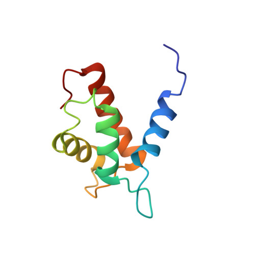
Solution structure of human Ca(2+)-bound S100A12

Hung, K.W.Hsu, C.C.Yu, C.

(2013) J Biomol NMR 57: 313-318

Macromolecule Content 

  • Total Structure Weight: 21.18 kDa 
  • Atom Count: 1,494 
  • Modeled Residue Count: 184 
  • Deposited Residue Count: 184 
  • Unique protein chains: 1

Macromolecules

Find similar proteins by:|  3D Structure
Entity ID: 1
MoleculeChains  Sequence LengthOrganismDetailsImage
Protein S100-A12
A, B
92Homo sapiensMutation(s): 0 
Gene Names: S100A12
UniProt & NIH Common Fund Data Resources
Find proteins for P80511 (Homo sapiens)
Explore P80511 
Go to UniProtKB:  P80511
PHAROS:  P80511
GTEx:  ENSG00000163221 
Entity Groups
Sequence Clusters30% Identity50% Identity70% Identity90% Identity95% Identity100% Identity
UniProt GroupP80511
Sequence Annotations
Expand
Reference Sequence

Experimental Data & Validation

Experimental Data

  • Method: SOLUTION NMR
  • Conformers Calculated: 200 
  • Conformers Submitted: 20 
  • Selection Criteria: structures with the lowest energy 

Structure Validation

View Full Validation Report


Entry History 

Deposition Data

Revision History  (Full details and data files)

  • Version 1.0: 2014-04-23
    Type: Initial release
  • Version 1.1: 2023-06-14
    Changes: Data collection, Database references, Other
  • Version 1.2: 2024-05-15
    Changes: Data collection, Database references