2M5V | pdb_00002m5v

Three-dimensional structure of human NLRP10/PYNOD pyrin domain


Experimental Data Snapshot

  • Method: SOLUTION NMR
  • Conformers Calculated: 100 
  • Conformers Submitted: 20 
  • Selection Criteria: structures with the lowest energy 

wwPDB Validation 3D Report Full Report

Validation slider image for 2M5V

This is version 1.4 of the entry. See complete history

Literature

Three-Dimensional Structure of Human NLRP10/PYNOD Pyrin Domain Reveals a Homotypic Interaction Site Distinct from Its Mouse Homologue.

Su, M.Y.Kuo, C.I.Chang, C.F.Chang, C.I.

(2013) PLoS One 8: e67843-e67843

  • DOI: https://doi.org/10.1371/journal.pone.0067843
  • Primary Citation Related Structures: 
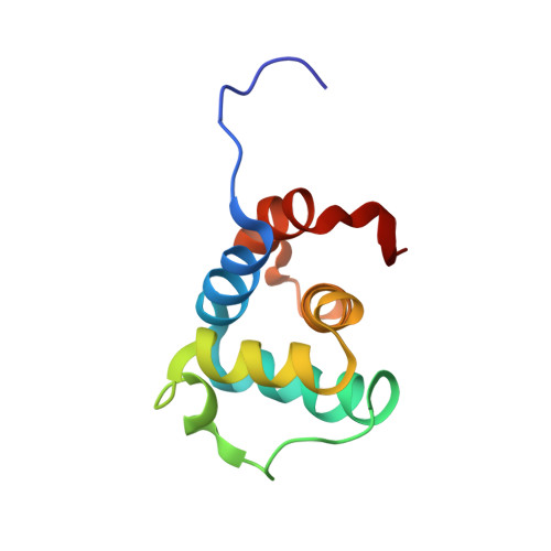
    2M5V

  • PubMed Abstract: 

    NLRPs (Nucleotide-binding domain, leucine-rich repeat and pyrin domain containing proteins) are a family of pattern-recognition receptors (PRRs) that sense intracellular microbial components and endogenous stress signals. NLRP10 (also known as PYNOD) is a unique NLRP member characterized by a lack of the putative ligand-binding leucine-rich repeat domain. Recently, human NLRP10 has been shown to inhibit the self-association of ASC into aggregates and ASC-mediated procaspase-1 processing. However, such activities are not found in mouse NLRP10. Here we report the solution structure and dynamics of human NLRP10 pyrin domain (PYD), whose helix H3 and loop H2-H3 adopt a conformation distinct from those of mouse NLRP10. Docking studies show that human and mouse NLRP10 PYDs may interact differently with ASC PYD. These results provide a possible structural explanation for the contrasting effect of NLRP10 on ASC aggregation in human cells versus mouse models. Finally, we also provide evidence that in human NLRP10 the PYD domain may not interact with the NOD domain to regulate its intrinsic nucleotide hydrolysis activity.


  • Organizational Affiliation
    • Institute of Biological Chemistry, Academia Sinica, Taipei, Taiwan, Republic of China.

Macromolecule Content 

  • Total Structure Weight: 11.58 kDa 
  • Atom Count: 810 
  • Modeled Residue Count: 102 
  • Deposited Residue Count: 102 
  • Unique protein chains: 1

Macromolecules

Find similar proteins by:|  3D Structure
Entity ID: 1
MoleculeChains  Sequence LengthOrganismDetailsImage
NACHT, LRR and PYD domains-containing protein 10102Homo sapiensMutation(s): 0 
Gene Names: NLRP10NALP10NOD8PYNOD
UniProt & NIH Common Fund Data Resources
Find proteins for Q86W26 (Homo sapiens)
Explore Q86W26 
Go to UniProtKB:  Q86W26
PHAROS:  Q86W26
GTEx:  ENSG00000182261 
Entity Groups
Sequence Clusters30% Identity50% Identity70% Identity90% Identity95% Identity100% Identity
UniProt GroupQ86W26
Sequence Annotations
Expand
Reference Sequence

Experimental Data & Validation

Experimental Data

  • Method: SOLUTION NMR
  • Conformers Calculated: 100 
  • Conformers Submitted: 20 
  • Selection Criteria: structures with the lowest energy 

Structure Validation

View Full Validation Report


Entry History 

Deposition Data

Revision History  (Full details and data files)

  • Version 1.0: 2013-04-03
    Type: Initial release
  • Version 1.1: 2013-06-05
    Changes: Database references
  • Version 1.2: 2013-07-31
    Changes: Database references
  • Version 1.3: 2023-06-14
    Changes: Data collection, Database references, Other
  • Version 1.4: 2024-05-15
    Changes: Data collection, Database references