2M55 | pdb_00002m55

NMR structure of the complex of an N-terminally acetylated alpha-synuclein peptide with calmodulin


Experimental Data Snapshot

  • Method: SOLUTION NMR
  • Conformers Calculated: 100 
  • Conformers Submitted: 20 
  • Selection Criteria: structures with the least restraint violations 

wwPDB Validation 3D Report Full Report

Validation slider image for 2M55

This is version 1.4 of the entry. See complete history

Literature

NMR Structure of Calmodulin Complexed to an N-Terminally Acetylated alpha-Synuclein Peptide.

Gruschus, J.M.Yap, T.L.Pistolesi, S.Maltsev, A.S.Lee, J.C.

(2013) Biochemistry 52: 3436-3445

  • DOI: https://doi.org/10.1021/bi400199p
  • Primary Citation Related Structures: 
    2M55

  • PubMed Abstract: 

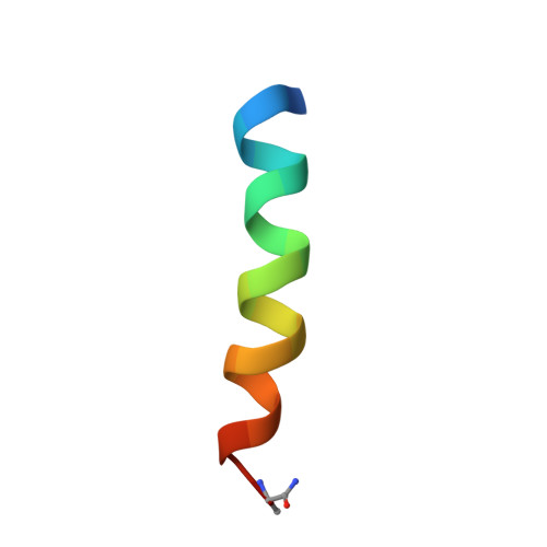
    Calmodulin (CaM) is a calcium binding protein that plays numerous roles in Ca-dependent cellular processes, including uptake and release of neurotransmitters in neurons. α-Synuclein (α-syn), one of the most abundant proteins in central nervous system neurons, helps maintain presynaptic vesicles containing neurotransmitters and moderates their Ca-dependent release into the synapse. Ca-Bound CaM interacts with α-syn most strongly at its N-terminus. The N-terminal region of α-syn is important for membrane binding; thus, CaM could modulate membrane association of α-syn in a Ca-dependent manner. In contrast, Ca-free CaM has negligible interaction. The interaction with CaM leads to significant signal broadening in both CaM and α-syn NMR spectra, most likely due to conformational exchange. The broadening is much reduced when binding a peptide consisting of the first 19 residues of α-syn. In neurons, most α-syn is acetylated at the N-terminus, and acetylation leads to a 10-fold increase in binding strength for the α-syn peptide (KD = 35 ± 10 μM). The N-terminally acetylated peptide adopts a helical structure at the N-terminus with the acetyl group contacting the N-terminal domain of CaM and with less ordered helical structure toward the C-terminus of the peptide contacting the CaM C-terminal domain. Comparison with known structures shows that the CaM/α-syn complex most closely resembles Ca-bound CaM in a complex with an IQ motif peptide. However, a search comparing the α-syn peptide sequence with known CaM targets, including IQ motifs, found no homologies; thus, the N-terminal α-syn CaM binding site appears to be a novel CaM target sequence.


  • Organizational Affiliation
    • Laboratory of Molecular Biophysics, Biochemistry and Biophysics Center, National Heart, Lung, and Blood Institute, National Institutes of Health , Bethesda, Maryland 20892, United States.

Macromolecule Content 

  • Total Structure Weight: 18.86 kDa 
  • Atom Count: 1,308 
  • Modeled Residue Count: 169 
  • Deposited Residue Count: 169 
  • Unique protein chains: 2

Macromolecules

Find similar proteins by:|  3D Structure
Entity ID: 1
MoleculeChains  Sequence LengthOrganismDetailsImage
Calmodulin148Homo sapiensMutation(s): 0 
Gene Names: 
CALM1CALMCAMCAM1CALM2CAM2CAMBCALM3CALML2CAM3...
CALM1CALMCAMCAM1CALM2CAM2CAMBCALM3CALML2CAM3CAMCCAMIII

UniProt & NIH Common Fund Data Resources
Find proteins for P0DP23 (Homo sapiens)
Explore P0DP23 
Go to UniProtKB:  P0DP23
PHAROS:  P0DP23
GTEx:  ENSG00000198668 
Entity Groups
Sequence Clusters30% Identity50% Identity70% Identity90% Identity95% Identity100% Identity
UniProt GroupP0DP23
Sequence Annotations
Expand
Reference Sequence
Find similar proteins by:  Sequence   |   3D Structure  
Entity ID: 2
MoleculeChains  Sequence LengthOrganismDetailsImage
Alpha-synuclein21Homo sapiensMutation(s): 2 
UniProt & NIH Common Fund Data Resources
Find proteins for P37840 (Homo sapiens)
Explore P37840 
Go to UniProtKB:  P37840
PHAROS:  P37840
GTEx:  ENSG00000145335 
Entity Groups
Sequence Clusters30% Identity50% Identity70% Identity90% Identity95% Identity100% Identity
UniProt GroupP37840
Sequence Annotations
Expand
Reference Sequence

Experimental Data & Validation

Experimental Data

  • Method: SOLUTION NMR
  • Conformers Calculated: 100 
  • Conformers Submitted: 20 
  • Selection Criteria: structures with the least restraint violations 

Structure Validation

View Full Validation Report


Entry History 

Deposition Data

Revision History  (Full details and data files)

  • Version 1.0: 2013-05-08
    Type: Initial release
  • Version 1.1: 2013-12-18
    Changes: Database references
  • Version 1.2: 2013-12-25
    Changes: Source and taxonomy
  • Version 1.3: 2023-06-14
    Changes: Data collection, Database references, Derived calculations, Other
  • Version 1.4: 2024-10-16
    Changes: Data collection, Database references, Structure summary