Impact of calcium binding and thionylation of S100A1 protein on its nuclear magnetic resonance-derived structure and backbone dynamics.
Nowakowski, M., Ruszczynska-Bartnik, K., Budzinska, M., Jaremko, L., Jaremko, M., Zdanowski, K., Bierzynski, A., Ejchart, A.(2013) Biochemistry 52: 1149-1159
- PubMed: 23351007 Search on PubMed
- DOI: https://doi.org/10.1021/bi3015407
- Primary Citation Related Structures:
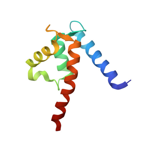
2LP2, 2LP3 - PubMed Abstract:
S100 proteins play a crucial role in multiple important biological processes in vertebrate organisms acting predominantly as calcium signal transmitters. S100A1 is a typical representative of this family of proteins. After four Ca(2+) ions bind, it undergoes a dramatic conformational change, resulting in exposure, in each of its two identical subunits, a large hydrophobic cleft that binds to target proteins. It has been shown that abnormal expression of S100A1 is strongly correlated with a number of severe human diseases: cardiomyopathy and neurodegenerative disorders. A few years ago, we found that thionylation of Cys 85, the unique cysteine in two identical S100A1 subunits, leads to a drastic increase of the affinity of the protein for calcium. We postulated that the protein activated by thionylation becomes a more efficient calcium signal transmitter. Therefore, we decided to undertake, using nuclear magnetic resonance methods, a comparative study of the structure and dynamics of native and thionylated human S100A1 in its apo and holo states. In this paper, we present the results obtained for both forms of this protein in its holo state and compare them with the previously published structure of native apo-S100. The main conclusion that we draw from these results is that the increased calcium binding affinity of S100A1 upon thionylation arises, most probably, from rearrangement of the hydrophobic core in its apo form.
- Institute of Biochemistry and Biophysics, Polish Academy of Sciences, Pawińskiego 5A, 02-106 Warsaw, Poland. lyam@chem.uw.edu.pl
Organizational Affiliation: