2LLQ | pdb_00002llq

Solution nmr-derived structure of calmodulin c-lobe bound with er alpha peptide


Experimental Data Snapshot

  • Method: SOLUTION NMR
  • Conformers Calculated: 200 
  • Conformers Submitted: 10 
  • Selection Criteria: structures with the lowest energy 

wwPDB Validation 3D Report Full Report

Validation slider image for 2LLQ

This is version 1.2 of the entry. See complete history

Literature

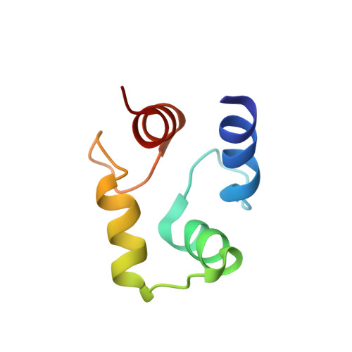
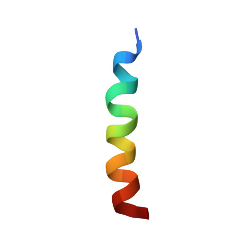
Structural basis for Ca2+-induced activation and dimerization of estrogen receptor alpha by calmodulin.

Zhang, Y.Li, Z.Sacks, D.B.Ames, J.B.

(2012) J Biological Chem 287: 9336-9344

  • DOI: https://doi.org/10.1074/jbc.M111.334797
  • Primary Citation Related Structures: 
    2LLO, 2LLQ

  • PubMed Abstract: 

    The estrogen receptor α (ER-α) regulates expression of target genes implicated in development, metabolism, and breast cancer. Calcium-dependent regulation of ER-α is critical for activating gene expression and is controlled by calmodulin (CaM). Here, we present the NMR structures for the two lobes of CaM each bound to a localized region of ER-α (residues 287-305). A model of the complete CaM·ER-α complex was constructed by combining these two structures with additional data. The two lobes of CaM both compete for binding at the same site on ER-α (residues 292, 296, 299, 302, and 303), which explains why full-length CaM binds two molecules of ER-α in a 1:2 complex and stabilizes ER-α dimerization. Exposed glutamate residues in CaM (Glu(11), Glu(14), Glu(84), and Glu(87)) form salt bridges with key lysine residues in ER-α (Lys(299), Lys(302), and Lys(303)), which are likely to prevent ubiquitination at these sites and inhibit degradation of ER-α. Mutants of ER-α at the CaM-binding site (W292A and K299A) weaken binding to CaM, and I298E/K299D disrupts estrogen-induced transcription. CaM facilitates dimerization of ER-α in the absence of estrogen, and stimulation of ER-α by either Ca(2+) and/or estrogen may serve to regulate transcription in a combinatorial fashion.


  • Organizational Affiliation
    • Department of Chemistry, University of California, Davis, California 95616, USA.

Macromolecule Content 

  • Total Structure Weight: 10.02 kDa 
  • Atom Count: 695 
  • Modeled Residue Count: 86 
  • Deposited Residue Count: 86 
  • Unique protein chains: 2

Macromolecules

Find similar proteins by:|  3D Structure
Entity ID: 1
MoleculeChains  Sequence LengthOrganismDetailsImage
Calmodulin67Xenopus laevisMutation(s): 0 
Gene Names: calm1calm2
UniProt
Find proteins for P0DP33 (Xenopus laevis)
Explore P0DP33 
Go to UniProtKB:  P0DP33
Entity Groups
Sequence Clusters30% Identity50% Identity70% Identity90% Identity95% Identity100% Identity
UniProt GroupP0DP33
Sequence Annotations
Expand
Reference Sequence
Find similar proteins by:  Sequence   |   3D Structure  
Entity ID: 2
MoleculeChains  Sequence LengthOrganismDetailsImage
Estrogen receptor19Homo sapiensMutation(s): 0 
UniProt & NIH Common Fund Data Resources
Find proteins for P03372 (Homo sapiens)
Explore P03372 
Go to UniProtKB:  P03372
PHAROS:  P03372
GTEx:  ENSG00000091831 
Entity Groups
Sequence Clusters30% Identity50% Identity70% Identity90% Identity95% Identity100% Identity
UniProt GroupP03372
Sequence Annotations
Expand
Reference Sequence

Small Molecules

Experimental Data & Validation

Experimental Data

  • Method: SOLUTION NMR
  • Conformers Calculated: 200 
  • Conformers Submitted: 10 
  • Selection Criteria: structures with the lowest energy 

Structure Validation

View Full Validation Report


Entry History 

Deposition Data

  • Released Date: 2012-02-01 
  • Deposition Author(s): Zhang, Y.

Revision History  (Full details and data files)

  • Version 1.0: 2012-02-01
    Type: Initial release
  • Version 1.1: 2013-08-28
    Changes: Database references
  • Version 1.2: 2024-05-01
    Changes: Data collection, Database references, Derived calculations