2L6Y | pdb_00002l6y

haddock model of GATA1NF:Lmo2LIM2-Ldb1LID


Experimental Data Snapshot

  • Method: SOLUTION NMR
  • Conformers Calculated: 10 
  • Conformers Submitted: 10 
  • Selection Criteria: all calculated structures submitted 

wwPDB Validation 3D Report Full Report

Validation slider image for 2L6Y

This is version 1.2 of the entry. See complete history

Literature

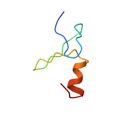
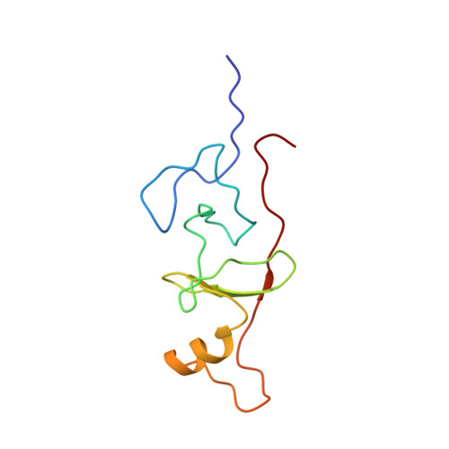
Structural basis of simultaneous recruitment of the transcriptional regulators LMO2 and FOG1/ZFPM1 by the transcription factor GATA1

Wilkinson-White, L.Gamsjaeger, R.Dastmalchi, S.Wienert, B.Stokes, P.H.Crossley, M.Mackay, J.P.Matthews, J.M.

(2011) Proc Natl Acad Sci U S A 108: 14443-14448

  • DOI: https://doi.org/10.1073/pnas.1105898108
  • Primary Citation Related Structures: 
    2L6Y, 2L6Z

  • PubMed Abstract: 

    The control of red blood cell and megakaryocyte development by the regulatory protein GATA1 is a paradigm for transcriptional regulation of gene expression in cell lineage differentiation and maturation. Most GATA1-regulated events require GATA1 to bind FOG1, and essentially all GATA1-activated genes are cooccupied by a TAL1/E2A/LMO2/LDB1 complex; however, it is not known whether FOG1 and TAL1/E2A/LMO2/LDB1 are simultaneously recruited by GATA1. Our structural data reveal that the FOG1-binding domain of GATA1, the N finger, can also directly contact LMO2 and show that, despite the small size (< 50 residues) of the GATA1 N finger, both FOG1 and LMO2 can simultaneously bind this domain. LMO2 in turn can simultaneously contact both GATA1 and the DNA-binding protein TAL1/E2A at bipartite E-box/WGATAR sites. Taken together, our data provide the first structural snapshot of multiprotein complex formation at GATA1-dependent genes and support a model in which FOG1 and TAL1/E2A/LMO2/LDB1 can cooccupy E-box/WGATAR sites to facilitate GATA1-mediated activation of gene activation.


  • Organizational Affiliation
    • School of Molecular Bioscience, University of Sydney, New South Wales, 2006 Sydney, Australia.

Macromolecule Content 

  • Total Structure Weight: 15.18 kDa 
  • Atom Count: 1,043 
  • Modeled Residue Count: 135 
  • Deposited Residue Count: 135 
  • Unique protein chains: 2

Macromolecules

Find similar proteins by:|  3D Structure
Entity ID: 1
MoleculeChains  Sequence LengthOrganismDetailsImage
Erythroid transcription factor39Mus musculusMutation(s): 0 
UniProt
Find proteins for P17679 (Mus musculus)
Explore P17679 
Go to UniProtKB:  P17679
Entity Groups
Sequence Clusters30% Identity50% Identity70% Identity90% Identity95% Identity100% Identity
UniProt GroupP17679
Sequence Annotations
Expand
Reference Sequence
Find similar proteins by:|  3D Structure
Entity ID: 2
MoleculeChains  Sequence LengthOrganismDetailsImage
LIM domain only 2, linker, LIM domain-binding protein 196Mus musculusMutation(s): 1 
UniProt
Find proteins for P25801 (Mus musculus)
Explore P25801 
Go to UniProtKB:  P25801
Find proteins for P70662 (Mus musculus)
Explore P70662 
Go to UniProtKB:  P70662
Entity Groups
Sequence Clusters30% Identity50% Identity70% Identity90% Identity95% Identity100% Identity
UniProt GroupsP70662P25801
Sequence Annotations
Expand
Reference Sequence

Experimental Data & Validation

Experimental Data

  • Method: SOLUTION NMR
  • Conformers Calculated: 10 
  • Conformers Submitted: 10 
  • Selection Criteria: all calculated structures submitted 

Structure Validation

View Full Validation Report


Entry History 

Deposition Data

Revision History  (Full details and data files)

  • Version 1.0: 2011-08-31
    Type: Initial release
  • Version 1.1: 2011-09-28
    Changes: Database references, Structure summary
  • Version 1.2: 2024-05-01
    Changes: Data collection, Database references, Derived calculations