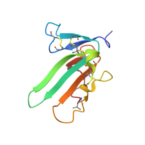
The TbetaR-I Pre-Helix Extension Is Structurally Ordered in the Unbound Form and Its Flanking Prolines Are Essential for Binding
Zuniga, J.E., Ilangovan, U., Mahlawat, P., Hinck, C.S., Huang, T., Groppe, J.C., McEwen, D.G., Hinck, A.P.(2011) J Mol Biology 412: 601-618
- PubMed: 21821041
- DOI: https://doi.org/10.1016/j.jmb.2011.07.046
- Primary Citation of Related Structures:
2L5S - PubMed Abstract:
Transforming growth factor β isoforms (TGF-β) are among the most recently evolved members of a signaling superfamily with more than 30 members. TGF-β play vital roles in regulating cellular growth and differentiation, and they signal through a highly restricted subset of receptors known as TGF-β type I receptor (TβR-I) and TGF-β type II receptor (TβR-II). TGF-β's specificity for TβR-I has been proposed to arise from its pre-helix extension, a five-residue loop that binds in the cleft between TGF-β and TβR-II. The structure and backbone dynamics of the unbound form of the TβR-I extracellular domain were determined using NMR to investigate the extension's role in binding. This showed that the unbound form is highly similar to the bound form in terms of both the β-strand framework that defines the three-finger toxin fold and the extension and its characteristic cis-Ile54-Pro55 peptide bond. The NMR data further showed that the extension and two flanking 3(10) helices are rigid on the nanosecond-to-picosecond timescale. The functional significance of several residues within the extension was investigated by binding studies and reporter gene assays in cultured epithelial cells. These demonstrated that the pre-helix extension is essential for binding, with Pro55 and Pro59 each playing a major role. These findings suggest that the pre-helix extension and its flanking prolines evolved to endow the TGF-β signaling complex with its unique specificity, departing from the ancestral promiscuity of the bone morphogenetic protein subfamily, where the binding interface of the type I receptor is highly flexible.
- Department of Biochemistry, University of Texas Health Science Center at San Antonio, San Antonio, TX 78229, USA.
Organizational Affiliation:


