2KZU | pdb_00002kzu

DAXX helical bundle (DHB) domain / Rassf1C complex


Experimental Data Snapshot

  • Method: SOLUTION NMR
  • Conformers Calculated: 120 
  • Conformers Submitted: 25 
  • Selection Criteria: structures with the lowest energy 

wwPDB Validation 3D Report Full Report

Validation slider image for 2KZU

This is version 1.4 of the entry. See complete history

Literature

Structural Characterization of the DAXX N-Terminal Helical Bundle Domain and Its Complex with Rassf1C.

Escobar-Cabrera, E.Lau, D.K.Giovinazzi, S.Ishov, A.M.McIntosh, L.P.

(2010) Structure 18: 1642-1653

  • DOI: https://doi.org/10.1016/j.str.2010.09.016
  • Primary Citation Related Structures: 
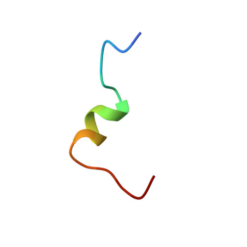
    2KZS, 2KZU

  • PubMed Abstract: 

    DAXX is a scaffold protein with diverse roles including transcription and cell cycle regulation. Using NMR spectroscopy, we demonstrate that the C-terminal half of DAXX is intrinsically disordered, whereas a folded domain is present near its N terminus. This domain forms a left-handed four-helix bundle (H1, H2, H4, H5). However, due to a crossover helix (H3), this topology differs from that of the Sin3 PAH domain, which to date has been used as a model for DAXX. The N-terminal residues of the tumor suppressor Rassf1C fold into an amphipathic α helix upon binding this DAXX domain via a shallow cleft along the flexible helices H2 and H5 (K(D) ∼60 μM). Based on a proposed DAXX recognition motif as hydrophobic residues preceded by negatively charged groups, we found that peptide models of p53 and Mdm2 also bound the helical bundle. These data provide a structural foundation for understanding the diverse functions of DAXX.


  • Organizational Affiliation
    • Department of Biochemistry and Molecular Biology, University of British Columbia, Vancouver, BC V6T1Z3, Canada.

Macromolecule Content 

  • Total Structure Weight: 13.1 kDa 
  • Atom Count: 919 
  • Modeled Residue Count: 112 
  • Deposited Residue Count: 112 
  • Unique protein chains: 2

Macromolecules

Find similar proteins by:|  3D Structure
Entity ID: 1
MoleculeChains  Sequence LengthOrganismDetailsImage
Death-associated protein 694Homo sapiensMutation(s): 0 
Gene Names: DAXXDADB-159G18.9-007DAMC-227D19.15-007DAQB-126H3.2-007XXbac-BCX165D10.3-007XXbac-BPG185D15.6-007
UniProt & NIH Common Fund Data Resources
Find proteins for Q9UER7 (Homo sapiens)
Explore Q9UER7 
Go to UniProtKB:  Q9UER7
PHAROS:  Q9UER7
GTEx:  ENSG00000204209 
Entity Groups
Sequence Clusters30% Identity50% Identity70% Identity90% Identity95% Identity100% Identity
UniProt GroupQ9UER7
Sequence Annotations
Expand
Reference Sequence
Find similar proteins by:  Sequence   |   3D Structure  
Entity ID: 2
MoleculeChains  Sequence LengthOrganismDetailsImage
Ras association (RalGDS/AF-6) domain family 118Homo sapiensMutation(s): 0 
Gene Names: RASSF1hCG_17462
UniProt & NIH Common Fund Data Resources
Find proteins for Q9NS23 (Homo sapiens)
Explore Q9NS23 
Go to UniProtKB:  Q9NS23
PHAROS:  Q9NS23
GTEx:  ENSG00000068028 
Entity Groups
Sequence Clusters30% Identity50% Identity70% Identity90% Identity95% Identity100% Identity
UniProt GroupQ9NS23
Sequence Annotations
Expand
Reference Sequence

Experimental Data & Validation

Experimental Data

  • Method: SOLUTION NMR
  • Conformers Calculated: 120 
  • Conformers Submitted: 25 
  • Selection Criteria: structures with the lowest energy 

Structure Validation

View Full Validation Report


Entry History 

Deposition Data

Revision History  (Full details and data files)

  • Version 1.0: 2010-12-15
    Type: Initial release
  • Version 1.1: 2011-07-13
    Changes: Version format compliance
  • Version 1.2: 2020-02-05
    Changes: Database references, Other
  • Version 1.3: 2023-06-14
    Changes: Database references, Other
  • Version 1.4: 2024-05-15
    Changes: Data collection, Database references