2KXF | pdb_00002kxf

Solution structure of the first two RRM domains of FBP-interacting repressor (FIR)


Experimental Data Snapshot

  • Method: SOLUTION NMR
  • Conformers Calculated: 200 
  • Conformers Submitted: 20 
  • Selection Criteria: structures with the lowest energy 

wwPDB Validation 3D Report Full Report

Validation slider image for 2KXF

This is version 1.3 of the entry. See complete history

Literature

Molecular basis of FIR-mediated c-myc transcriptional control.

Cukier, C.D.Hollingworth, D.Martin, S.R.Kelly, G.Diaz-Moreno, I.Ramos, A.

(2010) Nat Struct Mol Biol 17: 1058-1064

  • DOI: https://doi.org/10.1038/nsmb.1883
  • Primary Citation Related Structures: 
    2KXF, 2KXH

  • PubMed Abstract: 

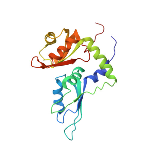
    The far upstream element (FUSE) regulatory system promotes a peak in the concentration of c-Myc during cell cycle. First, the FBP transcriptional activator binds to the FUSE DNA element upstream of the c-myc promoter. Then, FBP recruits its specific repressor (FIR), which acts as an on/off transcriptional switch. Here we describe the molecular basis of FIR recruitment, showing that the tandem RNA recognition motifs of FIR provide a platform for independent FUSE DNA and FBP protein binding and explaining the structural basis of the reversibility of the FBP-FIR interaction. We also show that the physical coupling between FBP and FIR is modulated by a flexible linker positioned sequentially to the recruiting element. Our data explain how the FUSE system precisely regulates c-myc transcription and suggest that a small change in FBP-FIR affinity leads to a substantial effect on c-Myc concentration.


  • Organizational Affiliation
    • Molecular Structure Division, Medical Research Council National Institute for Medical Research, London, UK.

Macromolecule Content 

  • Total Structure Weight: 21.93 kDa 
  • Atom Count: 1,538 
  • Modeled Residue Count: 199 
  • Deposited Residue Count: 199 
  • Unique protein chains: 1

Macromolecules

Find similar proteins by:|  3D Structure
Entity ID: 1
MoleculeChains  Sequence LengthOrganismDetailsImage
Poly(U)-binding-splicing factor PUF60199Homo sapiensMutation(s): 0 
UniProt & NIH Common Fund Data Resources
Find proteins for Q9UHX1 (Homo sapiens)
Explore Q9UHX1 
Go to UniProtKB:  Q9UHX1
PHAROS:  Q9UHX1
GTEx:  ENSG00000179950 
Entity Groups
Sequence Clusters30% Identity50% Identity70% Identity90% Identity95% Identity100% Identity
UniProt GroupQ9UHX1
Sequence Annotations
Expand
Reference Sequence

Experimental Data & Validation

Experimental Data

  • Method: SOLUTION NMR
  • Conformers Calculated: 200 
  • Conformers Submitted: 20 
  • Selection Criteria: structures with the lowest energy 

Structure Validation

View Full Validation Report


Entry History 

Deposition Data

Revision History  (Full details and data files)

  • Version 1.0: 2010-08-18
    Type: Initial release
  • Version 1.1: 2011-07-13
    Changes: Version format compliance
  • Version 1.2: 2019-12-18
    Changes: Data collection, Database references, Derived calculations, Experimental preparation
  • Version 1.3: 2024-05-01
    Changes: Data collection, Database references