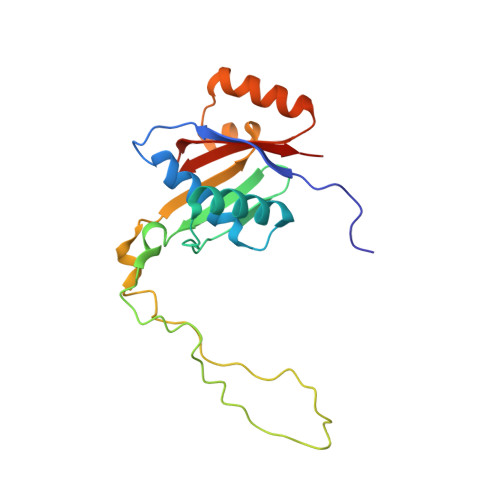
The binding mode of ATP revealed by the solution structure of the N-domain of human ATP7A.
Banci, L., Bertini, I., Cantini, F., Inagaki, S., Migliardi, M., Rosato, A.(2009) J Biological Chem
- PubMed: 19917612
- DOI: https://doi.org/10.1074/jbc.M109.054262
- Primary Citation of Related Structures:
2KMV, 2KMX - PubMed Abstract:
We report the solution NMR structures of the N-domain of the Menkes protein (ATP7A) in the ATP-free and ATP-bound forms. The structures consist of a twisted antiparallel six-stranded beta-sheet flanked by two pairs of alpha-helices. A protein loop of 50 amino acids located between beta 3 and beta 4 is disordered and mobile on the subnanosecond time scale. ATP binds with an affinity constant of (1.2 +/- 0.1) x 10(4) m(-1) and exchanges with a rate of the order of 1 x 10(3) s(-1). The ATP-binding cavity is considerably affected by the presence of the ligand, resulting in a more compact conformation in the ATP-bound than in the ATP-free form. This structural variation is due to the movement of the alpha1-alpha2 and beta2-beta 3 loops, both of which are highly conserved in copper(I)-transporting P(IB)-type ATPases. The present structure reveals a characteristic binding mode of ATP within the protein scaffold of the copper(I)-transporting P(IB)-type ATPases with respect to the other P-type ATPases. In particular, the binding cavity contains mainly hydrophobic aliphatic residues, which are involved in van der Waal's interactions with the adenine ring of ATP, and a Glu side chain, which forms a crucial hydrogen bond to the amino group of ATP.
- Magnetic Resonance Center (CERM) and Department of Chemistry, University of Florence, Italy.
Organizational Affiliation:


