Combined X-ray and NMR analysis of the stability of the cyclotide cystine knot fold that underpins its insecticidal activity and potential use as a drug scaffold
Wang, C.K., Hu, S.-H., Martin, J.L., Sjogren, T., Hajdu, J., Bohlin, L., Claeson, P., Goransson, U., Rosengren, K.J., Tang, J., Tan, N.-H., Craik, D.J.(2009) J Biol Chem 284: 10672-10683
- PubMed: 19211551 Search on PubMedSearch on PubMed Central
- DOI: https://doi.org/10.1074/jbc.M900021200
- Primary Citation Related Structures:
2K7G, 3E4H - PubMed Abstract:
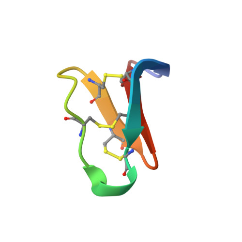
Cyclotides are a family of plant defense proteins that are highly resistant to adverse chemical, thermal, and enzymatic treatment. Here, we present the first crystal structure of a cyclotide, varv F, from the European field pansy, Viola arvensis, determined at a resolution of 1.8 A. The solution state NMR structure was also determined and, combined with measurements of biophysical parameters for several cyclotides, provided an insight into the structural features that account for the remarkable stability of the cyclotide family. The x-ray data confirm the cystine knot topology and the circular backbone, and delineate a conserved network of hydrogen bonds that contribute to the stability of the cyclotide fold. The structural role of a highly conserved Glu residue that has been shown to regulate cyclotide function was also determined, verifying its involvement in a stabilizing hydrogen bond network. We also demonstrate that varv F binds to dodecylphosphocholine micelles, defining the binding orientation and showing that its structure remains unchanged upon binding, further demonstrating that the cyclotide fold is rigid. This study provides a biological insight into the mechanism by which cyclotides maintain their native activity in the unfavorable environment of predator insect guts. It also provides a structural basis for explaining how a cluster of residues important for bioactivity may be involved in self-association interactions in membranes. As well as being important for their bioactivity, the structural rigidity of cyclotides makes them very suitable as a stable template for peptide-based drug design.
- University of Queensland, Institute for Molecular Bioscience, Brisbane, Queensland 4072, Australia.
Organizational Affiliation: