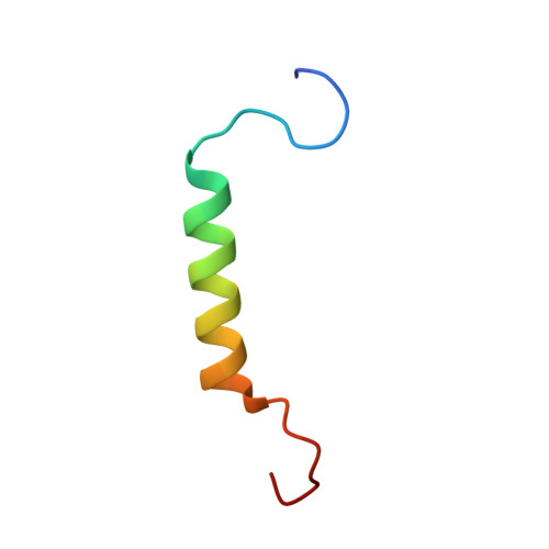
Structure of the first transmembrane domain of the neuronal acetylcholine receptor beta 2 subunit
Bondarenko, V., Xu, Y., Tang, P.(2007) Biophys J 92: 1616-1622
- PubMed: 17142275 Search on PubMedSearch on PubMed Central
- DOI: https://doi.org/10.1529/biophysj.106.095364
- Primary Citation Related Structures:
2K58 - PubMed Abstract:
The recent cryoelectron microscopy structure of the Torpedo nicotinic acetylcholine receptor (nAChR) at 4-A resolution shows long helices for all transmembrane (TM) domains. This is in disagreement with several previous reports that the first TM domain of nAChR and other Cys-loop receptors are not entirely helical. In this study, we determined the structure and backbone dynamics of an extended segment encompassing the first TM domain (TM1e) of nAChR beta(2) subunit in dodecylphosphocholine micelles, using solution-state NMR and circular dichroism (CD) spectroscopy. Both CD and NMR results show less helicity in TM1e than in Torpedo nAChR structure (Protein Data Bank: 2BG9). The helical ending residues at the C-terminus are the same in the TM1e NMR structure and the Torpedo nAChR structure, but the helical starting residue (I-217) in TM1e is seven residues closer to the C-terminus. Interestingly, the helical starting residue is two residues before the highly conserved P-219, in accordance with the hypothesis that proline causes helical distortions at three residues preceding it. The NMR relaxation measurements show a dynamics pattern consistent with TM1e structure. The substantial nonhelical content adds greater flexibilities to TM1e, thereby implicating a different molecular basis for nAChR function compared to a longer and more rigid helical TM1.
- Department of Anesthesiology, University of Pittsburgh School of Medicine, Pittsburgh, Pennsylvania 15260, USA.
Organizational Affiliation: