2K0F

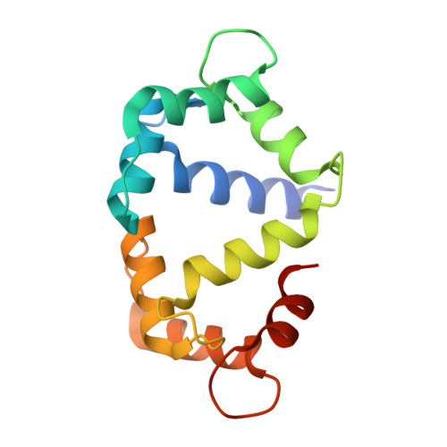
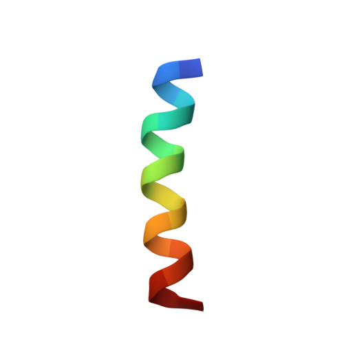
Calmodulin complexed with calmodulin-binding peptide from smooth muscle myosin light chain kinase


Experimental Data Snapshot

  • Method: SOLUTION NMR
  • Conformers Calculated: 160 
  • Conformers Submitted: 160 
  • Selection Criteria: all calculated structures submitted 

wwPDB Validation   3D Report Full Report


This is version 1.3 of the entry. See complete history


Literature

A coupled equilibrium shift mechanism in calmodulin-mediated signal transduction

Gsponer, J.Christodoulou, J.Cavalli, A.Bui, J.M.Richter, B.Dobson, C.M.Vendruscolo, M.

(2008) Structure 16: 736-746

  • DOI: https://doi.org/10.1016/j.str.2008.02.017
  • Primary Citation of Related Structures:  
    2K0E, 2K0F

  • PubMed Abstract: 

    We used nuclear magnetic resonance data to determine ensembles of conformations representing the structure and dynamics of calmodulin (CaM) in the calcium-bound state (Ca(2+)-CaM) and in the state bound to myosin light chain kinase (CaM-MLCK). These ensembles reveal that the Ca(2+)-CaM state includes a range of structures similar to those present when CaM is bound to MLCK. Detailed analysis of the ensembles demonstrates that correlated motions within the Ca(2+)-CaM state direct the structural fluctuations toward complex-like substates. This phenomenon enables initial ligation of MLCK at the C-terminal domain of CaM and induces a population shift among the substates accessible to the N-terminal domain, thus giving rise to the cooperativity associated with binding. Based on these results and the combination of modern free energy landscape theory with classical allostery models, we suggest that a coupled equilibrium shift mechanism controls the efficient binding of CaM to a wide range of ligands.


  • Organizational Affiliation

    Department of Chemistry, University of Cambridge, Lensfield Road, Cambridge CB2 1EW, United Kingdom.


Macromolecules
Find similar proteins by:  (by identity cutoff)  |  3D Structure
Entity ID: 1
MoleculeChains Sequence LengthOrganismDetailsImage
calmodulin148Homo sapiensMutation(s): 0 
Gene Names: CALM1CALMCAMCAM1
UniProt & NIH Common Fund Data Resources
Find proteins for P0DP23 (Homo sapiens)
Explore P0DP23 
Go to UniProtKB:  P0DP23
PHAROS:  P0DP23
GTEx:  ENSG00000198668 
Entity Groups  
Sequence Clusters30% Identity50% Identity70% Identity90% Identity95% Identity100% Identity
UniProt GroupP0DP23
Sequence Annotations
Expand
  • Reference Sequence

Find similar proteins by:  Sequence   |   3D Structure  

Entity ID: 2
MoleculeChains Sequence LengthOrganismDetailsImage
19-mer peptide from Myosin light chain kinase19N/AMutation(s): 1 
UniProt
Find proteins for P11799 (Gallus gallus)
Explore P11799 
Go to UniProtKB:  P11799
Entity Groups  
Sequence Clusters30% Identity50% Identity70% Identity90% Identity95% Identity100% Identity
UniProt GroupP11799
Sequence Annotations
Expand
  • Reference Sequence
Experimental Data & Validation

Experimental Data

  • Method: SOLUTION NMR
  • Conformers Calculated: 160 
  • Conformers Submitted: 160 
  • Selection Criteria: all calculated structures submitted 

Structure Validation

View Full Validation Report


Entry History 

Deposition Data

Revision History  (Full details and data files)

  • Version 1.0: 2008-06-10
    Type: Initial release
  • Version 1.1: 2011-07-13
    Changes: Version format compliance
  • Version 1.2: 2021-10-20
    Changes: Database references, Derived calculations
  • Version 1.3: 2024-05-29
    Changes: Data collection