Identification and Structural Characterization of an Unusual RING-Like Sequence within an Extracellular Biomineralization Protein, AP7.
Collino, S., Kim, I.W., Evans, J.S.(2008) Biochemistry 47: 3745-3755
- PubMed: 18298090 Search on PubMed
- DOI: https://doi.org/10.1021/bi701949p
- Primary Citation Related Structures:
2JYP - PubMed Abstract:
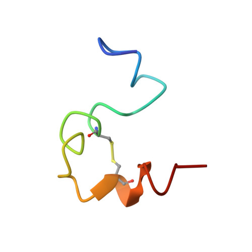
The RING or Really Interesting New Gene represents a family of eukaryotic sequences that bind Zn (II) ions and participate in intracellular processes involving protein-protein interaction. Although found in over 400 different proteins, very little is known regarding the structure-function properties of these domains because of the aggregation problems associated with RING sequences. To augment this data set, we report an unusual 36 AA C-terminal sequence of an extracellular matrix mollusk shell protein, AP7, that exhibits partial homology to the RING family. This Cys, His-containing sequence, termed AP7C, binds Zn (II) and other multivalent ions, but does not utilize a tetracoordinate complexation scheme for binding such as that found in Zn (II) finger polypeptides. Moreover, unlike Zn (II) finger and RING domains, this 36 AA can fold into a relatively stable structure in the absence of Zn (II). This folded structure consists of three short helical segments (A, B, and C), with segments A and B separated by a 4 AA type I beta-turn region and segments B and C separated by a 7 AA loop-like region. Interestingly, the putative RING-like region, -RRPFHECALCYSI-, experiences slow conformational exchange between two structural states in solution, most likely in response to imido ring interconversion at P8 and P21. Poisson-Boltzmann solvation calculations reveal that the AP7C molecular surface possesses a cationic region near its N-terminus, which lies adjacent to the 30 AA mineral modification domain in the AP7 protein. Given that the AP7C sequence does not influence mineralization, it is probable that this cationic pseudo-RING region is utilized by the AP7 protein for other tasks such as protein-protein interaction within the mollusk shell matrix.
- Laboratory for Chemical Physics, Center for Biomolecular Materials Spectroscopy, New York University, 345 E. 24th Street, Room 1007, New York, New York 10010, USA.
Organizational Affiliation: