Tryptophan Mutants of Cardiac Troponin C: 3D Structure, Troponin I Affinity, and in Situ Activity.
Julien, O., Sun, Y.B., Wang, X., Lindhout, D.A., Thiessen, A., Irving, M., Sykes, B.D.(2008) Biochemistry 47: 597-606
- PubMed: 18092822 Search on PubMed
- DOI: https://doi.org/10.1021/bi702056g
- Primary Citation Related Structures:
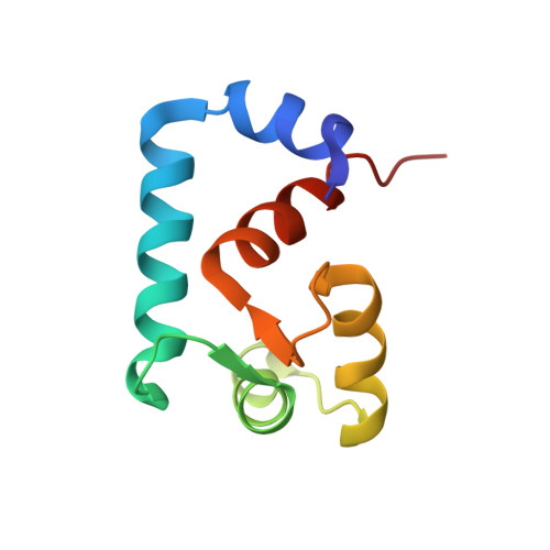
2JXL - PubMed Abstract:
In situ fluorescence/NMR spectroscopic approaches have been used to elucidate the structure, mobility, and domain orientations of troponin C in striated muscle. This led us to consider complementary approaches such as solid-state NMR spectroscopy. The biophysical properties of tryptophan and Trp-analogues, such as fluorotryptophan or hydroxytryptophan, are often exploited to probe protein structure and dynamics using solid-state NMR or fluorescence spectroscopy. We have characterized Phe-to-Trp mutants in the 'structural' C-domain of cardiac troponin C, designed to immobilize the indole ring in the hydrophobic core of the domain. The mutations and their fluorinated analogues (F104W, F104(5fW), F153W, and F153(5fW)) were shown not to perturb the structural properties of the protein. In this paper, we characterize the mutations F77W and F77W-V82A in the 'regulatory' N-domain of cardiac troponin C. We used NMR to determine the structure and dynamics of the mutant F77W-V82A-cNTnC, which shows a unique orientation of the indole ring. We observed a decrease in calcium binding affinity and a weaker affinity for the switch region of TnI for both mutants. We present force recovery measurements for all of the N- and C-domain mutants reconstituted into skeletal muscle fibers. The F77W mutation leads to a reduction of the in situ force recovery, whereas the C-domain mutants have the same activity as the wild type. These results suggest that the perturbations of the N-domain caused by the Trp mutation disturb the interaction between TnC and TnI, which in turn diminishes the activity in fibers, providing a clear example of the correlation between in vitro protein structures, their interactions, and the resulting in situ physiological activity.
- Department of Biochemistry, University of Alberta, Edmonton, Canada.
Organizational Affiliation: