Structural analysis of immunotherapeutic peptides for autoimmune Myasthenia gravis
Jung, H.H., Yi, H.J., Lee, S.K., Lee, J.Y., Jung, H.J., Yang, S.T., Eu, Y.-J., Im, S.-H., Kim, J.I.(2007) Biochemistry 46: 14987-14995
- PubMed: 18052043 Search on PubMed
- DOI: https://doi.org/10.1021/bi701298b
- Primary Citation Related Structures:
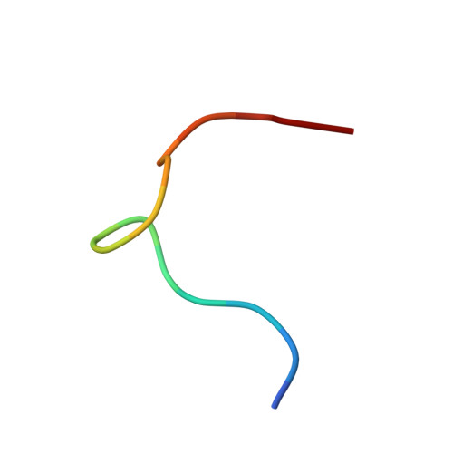
2JRV, 2JRW - PubMed Abstract:
Myasthenia gravis (MG) and its animal model, experimental MG (EAMG), are autoimmune disorders in which major pathogenic antibodies are directed against the main immunogenic region (MIR) of the nicotinic acetylcholine receptor (nAChR). In an earlier attempt to develop peptide mimotopes capable of preventing the anti-MIR-mediated pathogenicity, the peptide Pep.1 was initially identified from phage display, and subsequently, Cyclic extended Pep.1 (Cyc.ext.Pep.1), which incorporates eight additional residues into the Pep.1 sequence and has an affinity for the anti-MIR antibody mAb198 3 orders of magnitude greater than that of Pep.1, was developed. In an animal model, Pep.1 shows no ability to inhibit mAb198-induced EAMG, whereas Cyc.ext.Pep.1 successfully blocks anti-MIR antibody 198 (mAb198)-induced EAMG. Our aim in this study was to identify the structural characteristics related to the different affinities for mAb198 of Pep.1 and Cyc.ext.Pep.1 using NMR spectroscopy and alanine scanning analysis. The NMR structural analysis revealed that Pep.1 is very flexible in solution, whereas Cyc.ext.Pep.1 is highly rigid within a region containing several turn structures. Interestingly, TRNOE experiments revealed that mAb198-bound Pep.1, particularly in the region between Asn7 and Glu11, shows significant structural similarity to the region between Asn10 and Glu14 of Cyc.ext.Pep.1, which is critical for interaction with mAb198. We therefore conclude the higher affinity of Cyc.ext.Pep.1 for mAb198 reflects the fact that incorporation of additional residues producing a single disulfide bond endows Pep.1 with a conformational rigidity that mimics the structure of mAb198-bound Pep.1. Furthermore, our results suggest that cyclic extended peptides could be utilized generally as useful tools to optimize the affinity of phage library-derived peptide antigens.
- Department of Life Sciences, Research Center for Biomolecular Nanotechnology, Gwangju Institute of Science and Technology, Gwangju 500-712, Korea.
Organizational Affiliation: