2JPH | pdb_00002jph

NMR solution structure of the Rho GTPase binding domain of human plexin-b1


Experimental Data Snapshot

  • Method: SOLUTION NMR
  • Conformers Calculated: 100 
  • Conformers Submitted: 20 
  • Selection Criteria: structures with the least restraint violations 

wwPDB Validation 3D Report Full Report

Validation slider image for 2JPH

This is version 1.3 of the entry. See complete history

Literature

Insights into Oncogenic Mutations of Plexin-B1 Based on the Solution Structure of the Rho GTPase Binding Domain

Tong, Y.Hota, P.K.Hamaneh, M.B.Buck, M.

(2008) Structure 16: 246-258

  • DOI: https://doi.org/10.1016/j.str.2007.12.012
  • Primary Citation Related Structures: 
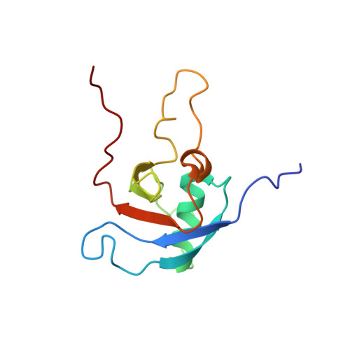
    2JPH

  • PubMed Abstract: 

    The plexin family of transmembrane receptors are important for axon guidance, angiogenesis, but also in cancer. Recently, plexin-B1 somatic missense mutations were found in both primary tumors and metastases of breast and prostate cancers, with several mutations mapping to the Rho GTPase binding domain (RBD) in the cytoplasmic region of the receptor. Here we present the NMR solution structure of this domain, confirming that the protein has both a ubiquitin-like fold and surface features. Oncogenic mutations T1795A and T1802A are located in a loop region, perturb the average structure locally, and have no effect on Rho GTPase binding affinity. Mutations L1815F and L1815P are located at the Rho GTPase binding site and are associated with a complete loss of binding for Rac1 and Rnd1. Both are found to disturb the conformation of the beta3-beta4 sheet and the orientation of surrounding side chains. Our study suggests that the oncogenic behavior of the mutants can be rationalized with reference to the structure of the RBD of plexin-B1.


  • Organizational Affiliation
    • Department of Physiology and Biophysics, Case Western Reserve University School of Medicine, 10900 Euclid Avenue, Cleveland, Ohio 44106, USA.

Macromolecule Content 

  • Total Structure Weight: 13.59 kDa 
  • Atom Count: 945 
  • Modeled Residue Count: 122 
  • Deposited Residue Count: 123 
  • Unique protein chains: 1

Macromolecules

Find similar proteins by:|  3D Structure
Entity ID: 1
MoleculeChains  Sequence LengthOrganismDetailsImage
Plexin-B1123Homo sapiensMutation(s): 1 
Gene Names: PLXNB1KIAA0407SEP
UniProt & NIH Common Fund Data Resources
Find proteins for O43157 (Homo sapiens)
Explore O43157 
Go to UniProtKB:  O43157
PHAROS:  O43157
GTEx:  ENSG00000164050 
Entity Groups
Sequence Clusters30% Identity50% Identity70% Identity90% Identity95% Identity100% Identity
UniProt GroupO43157
Sequence Annotations
Expand
Reference Sequence

Experimental Data & Validation

Experimental Data

  • Method: SOLUTION NMR
  • Conformers Calculated: 100 
  • Conformers Submitted: 20 
  • Selection Criteria: structures with the least restraint violations 

Structure Validation

View Full Validation Report


Entry History 

Deposition Data

Revision History  (Full details and data files)

  • Version 1.0: 2008-03-18
    Type: Initial release
  • Version 1.1: 2011-07-13
    Changes: Version format compliance
  • Version 1.2: 2021-10-20
    Changes: Data collection, Database references, Derived calculations
  • Version 1.3: 2023-12-20
    Changes: Data collection, Other