2JKG

Plasmodium falciparum profilin


Experimental Data Snapshot

  • Method: X-RAY DIFFRACTION
  • Resolution: 1.89 Å
  • R-Value Free: 0.217 
  • R-Value Work: 0.176 
  • R-Value Observed: 0.178 

wwPDB Validation   3D Report Full Report


This is version 1.4 of the entry. See complete history


Literature

Structural Basis for Parasite-Specific Functions of the Divergent Profilin of Plasmodium Falciparum.

Kursula, I.Kursula, P.Ganter, M.Panjikar, S.Matuschewski, K.Schuler, H.

(2008) Structure 16: 1638

  • DOI: https://doi.org/10.1016/j.str.2008.09.008
  • Primary Citation of Related Structures:  
    2JKF, 2JKG

  • PubMed Abstract: 

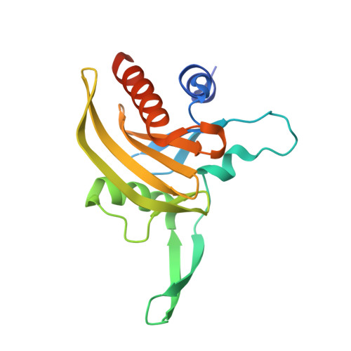
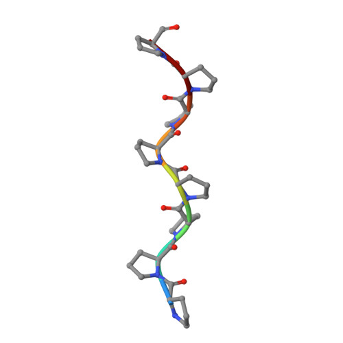
    Profilins are key regulators of actin dynamics. They sequester actin monomers, forming a pool for rapid polymer formation stimulated by proteins such as formins. Apicomplexan parasites utilize a highly specialized microfilament system for motility and host cell invasion. Their genomes encode only a small number of divergent actin regulators. We present the first crystal structure of an apicomplexan profilin, that of the malaria parasite Plasmodium falciparum, alone and in complex with a polyproline ligand peptide. The most striking feature of Plasmodium profilin is a unique minidomain consisting of a large beta-hairpin extension common to all apicomplexan parasites, and an acidic loop specific for Plasmodium species. Reverse genetics in the rodent malaria model, Plasmodium berghei, suggests that profilin is essential for the invasive blood stages of the parasite. Together, our data establish the structural basis for understanding the functions of profilin in the malaria parasite.


  • Organizational Affiliation

    Department of Biochemistry, University of Oulu, 90570 Oulu, Finland. inari.kursula@oulu.fi


Macromolecules
Find similar proteins by:  (by identity cutoff)  |  3D Structure
Entity ID: 1
MoleculeChains Sequence LengthOrganismDetailsImage
PROFILIN179Plasmodium falciparumMutation(s): 0 
UniProt
Find proteins for Q8I2J4 (Plasmodium falciparum (isolate 3D7))
Explore Q8I2J4 
Go to UniProtKB:  Q8I2J4
Entity Groups  
Sequence Clusters30% Identity50% Identity70% Identity90% Identity95% Identity100% Identity
UniProt GroupQ8I2J4
Sequence Annotations
Expand
  • Reference Sequence

Find similar proteins by:  Sequence   |   3D Structure  

Entity ID: 2
MoleculeChains Sequence LengthOrganismDetailsImage
OCTAPROLINE PEPTIDEB [auth P]8synthetic constructMutation(s): 0 
Sequence Annotations
Expand
  • Reference Sequence
Small Molecules
Ligands 1 Unique
IDChains Name / Formula / InChI Key2D Diagram3D Interactions
MG
Query on MG

Download Ideal Coordinates CCD File 
C [auth A]MAGNESIUM ION
Mg
JLVVSXFLKOJNIY-UHFFFAOYSA-N
Experimental Data & Validation

Experimental Data

  • Method: X-RAY DIFFRACTION
  • Resolution: 1.89 Å
  • R-Value Free: 0.217 
  • R-Value Work: 0.176 
  • R-Value Observed: 0.178 
  • Space Group: P 21 21 21
Unit Cell:
Length ( Å )Angle ( ˚ )
a = 45.52α = 90
b = 49.7β = 90
c = 83.81γ = 90
Software Package:
Software NamePurpose
REFMACrefinement
XDSdata reduction
XSCALEdata scaling
Auto-Rickshawphasing

Structure Validation

View Full Validation Report


Entry History 

Deposition Data

Revision History  (Full details and data files)

  • Version 1.0: 2008-09-23
    Type: Initial release
  • Version 1.1: 2011-07-13
    Changes: Advisory, Version format compliance
  • Version 1.2: 2016-12-21
    Changes: Data collection, Source and taxonomy, Structure summary
  • Version 1.3: 2018-01-17
    Changes: Data collection
  • Version 1.4: 2019-07-17
    Changes: Data collection