2JJL | pdb_00002jjl

Structure of avian reovirus sigmaC117-326, P321 crystal form


Experimental Data Snapshot

  • Method: X-RAY DIFFRACTION
  • Resolution: 2.30 Å
  • R-Value Free: 
    0.220 (Depositor), 0.217 (DCC) 
  • R-Value Work: 
    0.181 (Depositor), 0.177 (DCC) 
  • R-Value Observed: 
    0.184 (Depositor) 

Starting Model: experimental
View more details

wwPDB Validation 3D Report Full Report

Validation slider image for 2JJL

This is version 1.4 of the entry. See complete history

Literature

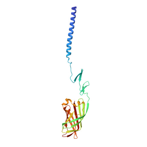
Crystallographic structure of the alpha-helical triple coiled-coil domain of avian reovirus S1133 fibre.

Guardado-Calvo, P.Fox, G.C.Llamas-Saiz, A.L.van Raaij, M.J.

(2009) J Gen Virol 90: 672-677

  • DOI: https://doi.org/10.1099/vir.0.008276-0
  • Primary Citation Related Structures: 
    2JJL, 2VRS

  • PubMed Abstract: 

    Avian reovirus fibre, a homo-trimer of the sigmaC protein, is a minor component of the avian reovirus outer capsid. It is anchored via a short N-terminal sequence to the inner capsid lambdaC pentamer, and its protruding globular C-terminal domain is responsible for primary host cell attachment. We have previously solved the structure of a receptor-binding fragment in which residues 160-191 form a triple beta-spiral and 196-326 a beta-barrel head domain. Here we have expressed, purified and crystallized a major sigmaC fragment comprising residues 117-326. Its structure, which was solved by molecular replacement using the previously determined receptor-binding domain structure and refined to 1.75 A (0.175 nm) resolution, reveals an alpha-helical triple coiled-coil connected to the previously solved structure by a zinc-ion-containing linker. The coiled-coil domain contains two chloride ion binding sites, as well as specific trimerization and registration sequences. The linker may act as a functionally important hinge.


  • Organizational Affiliation
    • Departamento de Bioquímica e Bioloxía Molecular, Facultade de Farmacia, Universidade de Santiago de Compostela, E-15782 Santiago de Compostela, Spain.

Macromolecule Content 

  • Total Structure Weight: 22.95 kDa 
  • Atom Count: 1,762 
  • Modeled Residue Count: 207 
  • Deposited Residue Count: 211 
  • Unique protein chains: 1

Macromolecules

Find similar proteins by:|  3D Structure
Entity ID: 1
MoleculeChains  Sequence LengthOrganismDetailsImage
SIGMA-C CAPSID PROTEIN211Avian orthoreovirusMutation(s): 0 
UniProt
Find proteins for Q992I2 (Avian reovirus (strain S1133))
Explore Q992I2 
Go to UniProtKB:  Q992I2
Entity Groups
Sequence Clusters30% Identity50% Identity70% Identity90% Identity95% Identity100% Identity
UniProt GroupQ992I2
Sequence Annotations
Expand
Reference Sequence

Experimental Data & Validation

Experimental Data

  • Method: X-RAY DIFFRACTION
  • Resolution: 2.30 Å
  • R-Value Free:  0.220 (Depositor), 0.217 (DCC) 
  • R-Value Work:  0.181 (Depositor), 0.177 (DCC) 
  • R-Value Observed: 0.184 (Depositor) 
Space Group: P 3 2 1
Unit Cell:
Length ( Å )Angle ( ˚ )
a = 77.657α = 90
b = 77.657β = 90
c = 121.449γ = 120
Software Package:
Software NamePurpose
REFMACrefinement
HKL-2000data reduction
SCALEPACKdata scaling
MOLREPphasing

Structure Validation

View Full Validation Report


Entry History 

Deposition Data

Revision History  (Full details and data files)

  • Version 1.0: 2009-01-13
    Type: Initial release
  • Version 1.1: 2011-05-07
    Changes: Version format compliance
  • Version 1.2: 2011-07-13
    Changes: Version format compliance
  • Version 1.3: 2018-02-07
    Changes: Database references
  • Version 1.4: 2023-12-13
    Changes: Data collection, Database references, Derived calculations, Other, Refinement description