2JIU | pdb_00002jiu

Crystal structure of EGFR kinase domain T790M mutation in complex with AEE788


Experimental Data Snapshot

  • Method: X-RAY DIFFRACTION
  • Resolution: 3.05 Å
  • R-Value Free: 
    0.277 (Depositor), 0.274 (DCC) 
  • R-Value Work: 
    0.211 (Depositor), 0.206 (DCC) 
  • R-Value Observed: 
    0.214 (Depositor) 

Starting Model: experimental
View more details

wwPDB Validation 3D Report Full Report

Validation slider image for 2JIU

Ligand Structure Quality Assessment 


This is version 1.3 of the entry. See complete history

Literature

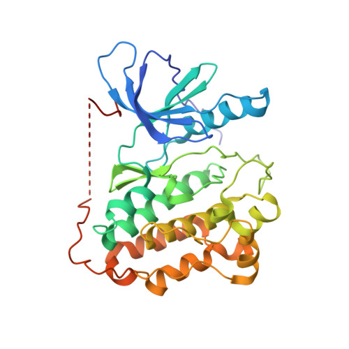
The T790M Mutation in Egfr Kinase Causes Drug Resistance by Increasing the Affinity for ATP.

Yun, C.-H.Mengwasser, K.E.Toms, A.V.Woo, M.S.Greulich, H.Wong, K.-K.Meyerson, M.Eck, M.J.

(2008) Proc Natl Acad Sci U S A 105: 2070

  • DOI: https://doi.org/10.1073/pnas.0709662105
  • Primary Citation Related Structures: 
    2JIT, 2JIU, 2JIV

  • PubMed Abstract: 

    Lung cancers caused by activating mutations in the epidermal growth factor receptor (EGFR) are initially responsive to small molecule tyrosine kinase inhibitors (TKIs), but the efficacy of these agents is often limited because of the emergence of drug resistance conferred by a second mutation, T790M. Threonine 790 is the "gatekeeper" residue, an important determinant of inhibitor specificity in the ATP binding pocket. The T790M mutation has been thought to cause resistance by sterically blocking binding of TKIs such as gefitinib and erlotinib, but this explanation is difficult to reconcile with the fact that it remains sensitive to structurally similar irreversible inhibitors. Here, we show by using a direct binding assay that T790M mutants retain low-nanomolar affinity for gefitinib. Furthermore, we show that the T790M mutation activates WT EGFR and that introduction of the T790M mutation increases the ATP affinity of the oncogenic L858R mutant by more than an order of magnitude. The increased ATP affinity is the primary mechanism by which the T790M mutation confers drug resistance. Crystallographic analysis of the T790M mutant shows how it can adapt to accommodate tight binding of diverse inhibitors, including the irreversible inhibitor HKI-272, and also suggests a structural mechanism for catalytic activation. We conclude that the T790M mutation is a "generic" resistance mutation that will reduce the potency of any ATP-competitive kinase inhibitor and that irreversible inhibitors overcome this resistance simply through covalent binding, not as a result of an alternative binding mode.


  • Organizational Affiliation
    • Department of Biological Chemistry and Molecular Pharmacology, Harvard Medical School, 25 Shattuck Street, Boston, MA 02115, USA.

Macromolecule Content 

  • Total Structure Weight: 75.28 kDa 
  • Atom Count: 4,880 
  • Modeled Residue Count: 614 
  • Deposited Residue Count: 656 
  • Unique protein chains: 1

Macromolecules

Find similar proteins by:|  3D Structure
Entity ID: 1
MoleculeChains  Sequence LengthOrganismDetailsImage
EPIDERMAL GROWTH FACTOR RECEPTOR
A, B
328Homo sapiensMutation(s): 1 
EC: 2.7.1.112 (PDB Primary Data), 2.7.10.1 (UniProt)
UniProt & NIH Common Fund Data Resources
Find proteins for P00533 (Homo sapiens)
Explore P00533 
Go to UniProtKB:  P00533
PHAROS:  P00533
GTEx:  ENSG00000146648 
Entity Groups
Sequence Clusters30% Identity50% Identity70% Identity90% Identity95% Identity100% Identity
UniProt GroupP00533
Sequence Annotations
Expand
Reference Sequence

Small Molecules

Ligands 1 Unique
IDChains Name / Formula / InChI Key2D Diagram3D Interactions
AEE

Query on AEE


Download:Ideal Coordinates CCD File
C [auth A]6-{4-[(4-ETHYLPIPERAZIN-1-YL)METHYL]PHENYL}-N-[(1R)-1-PHENYLETHYL]-7H-PYRROLO[2,3-D]PYRIMIDIN-4-AMINE
C27 H32 N6
OONFNUWBHFSNBT-HXUWFJFHSA-N

Experimental Data & Validation

Experimental Data

  • Method: X-RAY DIFFRACTION
  • Resolution: 3.05 Å
  • R-Value Free:  0.277 (Depositor), 0.274 (DCC) 
  • R-Value Work:  0.211 (Depositor), 0.206 (DCC) 
  • R-Value Observed: 0.214 (Depositor) 
Space Group: P 21 21 21
Unit Cell:
Length ( Å )Angle ( ˚ )
a = 48.405α = 90
b = 88.643β = 90
c = 164.867γ = 90
Software Package:
Software NamePurpose
REFMACrefinement
HKL-2000data reduction
HKL-2000data scaling
PHASERphasing

Structure Validation

View Full Validation Report


Ligand Structure Quality Assessment 


Entry History 

Deposition Data

Revision History  (Full details and data files)

  • Version 1.0: 2008-01-22
    Type: Initial release
  • Version 1.1: 2011-05-07
    Changes: Version format compliance
  • Version 1.2: 2011-07-13
    Changes: Version format compliance
  • Version 1.3: 2023-12-13
    Changes: Data collection, Database references, Other, Refinement description