Three-Dimensional Structure of a Putative Non- Cellulosomal Cohesin Module from a Clostridium Perfringens Family 84 Glycoside Hydrolase.
Chitayat, S., Gregg, K., Adams, J.J., Ficko-Blean, E., Bayer, E.A., Boraston, A.B., Smith, S.P.(2008) J Mol Biology 375: 20
- PubMed: 17999932 Search on PubMed
- DOI: https://doi.org/10.1016/j.jmb.2007.10.031
- Primary Citation Related Structures:
2JH2, 2O4E - PubMed Abstract:
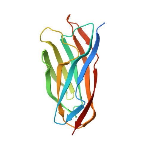
The genomes of myonecrotic strains of Clostridium perfringens encode a large number of secreted glycoside hydrolases. The activities of these enzymes are consistent with degradation of the mucosal layer of the human gastrointestinal tract, glycosaminoglycans and other cellular glycans found throughout the body. In many cases this is thought to aid in the propagation of the major toxins produced by C. perfringens. One such example is the family 84 glycoside hydrolases, which contains five C. perfringens members (CpGH84A-E), each displaying a unique modular architecture. The smallest and most extensively studied member, CpGH84C, comprises an N-terminal catalytic domain with beta-N-acetylglucosaminidase activity, a family 32 carbohydrate-binding module, a family 82 X-module (X82) of unknown function, and a fibronectin type-III-like module. Here we present the structure of the X82 module from CpGH84C, determined by both NMR spectroscopy and X-ray crystallography. CpGH84C X82 adopts a jell-roll fold comprising two beta-sheets formed by nine beta-strands. CpGH84C X82 displays distant amino acid sequence identity yet close structural similarity to the cohesin modules of cellulolytic anaerobic bacteria. Cohesin modules are responsible for the assembly of numerous hydrolytic enzymes in a cellulose-degrading multi-enzyme complex, termed the cellulosome, through a high-affinity interaction with the calcium-binding dockerin module. A planar surface is located on the face of the CpGH84 X82 structure that corresponds to the dockerin-binding region of cellulolytic cohesin modules and has the approximate dimensions to accommodate a dockerin module. The presence of cohesin-like X82 modules in glycoside hydrolases of C. perfringens is an indication that the formation of novel X82-dockerin mediated multi-enzyme complexes, with potential roles in pathogenesis, is possible.
- Department of Biochemistry, Queen's University, Kingston, Ontario, Canada.
Organizational Affiliation: