2JE4 | pdb_00002je4

Atomic-resolution crystal structure of chemically-synthesized HIV-1 protease in complex with JG-365


Experimental Data Snapshot

  • Method: X-RAY DIFFRACTION
  • Resolution: 1.07 Å
  • R-Value Free: 
    0.184 (Depositor) 
  • R-Value Observed: 
    0.143 (Depositor) 

Starting Model: experimental
View more details

wwPDB Validation 3D Report Full Report

Validation slider image for 2JE4

This is version 2.1 of the entry. See complete history

Literature

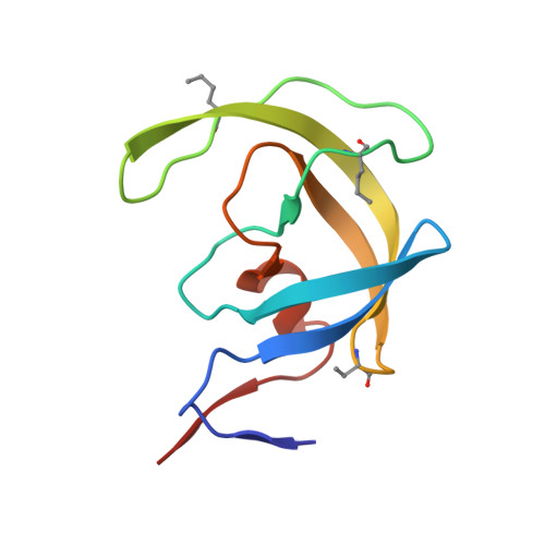
Modular Total Chemical Synthesis of a Human Immunodeficiency Virus Type 1 Protease.

Johnson, E.C.B.Malito, E.Shen, Y.Rich, D.Tang, W.J.Kent, S.B.

(2007) J Am Chem Soc 129: 11480

  • DOI: https://doi.org/10.1021/ja072870n
  • Primary Citation Related Structures: 
    2JE4

  • PubMed Abstract: 

    As part of our ongoing studies of the human immunodeficiency virus type 1 (HIV-1) protease enzyme, we set out to develop a modular chemical synthesis of the protein from multiple peptide segments. Our initial attempts were frustrated by the insolubility of intermediate peptide products. To overcome this problem, we designed a synthetic strategy combining the solubility-enhancing properties of C-terminal (Arg)n tags and the biological phenomenon of autoprocessing of the Gag-Pol polyprotein that occurs during maturation of the HIV-1 virus in vivo. Synthesis of a 119-residue peptide chain containing 10 residues of the reverse transcriptase (RT) open reading frame plus an (Arg)(10) tag at the C-terminus was straightforward by native chemical ligation followed by conversion of the Cys residues to Ala by Raney nickel desulfurization. The product polypeptide itself completed the final synthetic step by removing the C-terminal modification under folding conditions, to give the mature 99-residue polypeptide. High-purity homodimeric HIV-1 protease protein was obtained in excellent yield and had full enzymatic activity; the structure of the synthetic enzyme was confirmed by X-ray crystallography to a resolution of 1.07 A. This efficient modular synthesis by a biomimetic autoprocessing strategy will enable the facile synthesis of unique chemical analogues of the HIV-1 protease to further elucidate the molecular basis of enzyme catalysis.


  • Organizational Affiliation
    • Department of Biochemistry and Molecular Biology, Institute for Biophysical Dynamics, Ben-May Department for Cancer Research, The University of Chicago, Chicago, Illinois 60637, USA.

Macromolecule Content 

  • Total Structure Weight: 23.33 kDa 
  • Atom Count: 2,121 
  • Modeled Residue Count: 204 
  • Deposited Residue Count: 205 
  • Unique protein chains: 2

Macromolecules

Find similar proteins by:|  3D Structure
Entity ID: 1
MoleculeChains  Sequence LengthOrganismDetailsImage
PROTEASE
A, B
99Human immunodeficiency virus 1Mutation(s): 0 
UniProt
Find proteins for P03369 (Human immunodeficiency virus type 1 group M subtype B (isolate ARV2/SF2))
Explore P03369 
Go to UniProtKB:  P03369
Entity Groups
Sequence Clusters30% Identity50% Identity70% Identity90% Identity95% Identity100% Identity
UniProt GroupP03369
Sequence Annotations
Expand
Reference Sequence
Find similar proteins by:  Sequence   |   3D Structure  
Entity ID: 2
MoleculeChains  Sequence LengthOrganismDetailsImage
INHIBITOR MOLECULE JG3657synthetic constructMutation(s): 0 

Small Molecules

Ligands 3 Unique
IDChains Name / Formula / InChI Key2D Diagram3D Interactions
SO4

Query on SO4


Download:Ideal Coordinates CCD File
I [auth A],
J [auth A],
K [auth A],
N [auth B],
O [auth B]
SULFATE ION
O4 S
QAOWNCQODCNURD-UHFFFAOYSA-L
GOL

Query on GOL


Download:Ideal Coordinates CCD File
F [auth A],
G [auth A],
H [auth A]
GLYCEROL
C3 H8 O3
PEDCQBHIVMGVHV-UHFFFAOYSA-N
ACT

Query on ACT


Download:Ideal Coordinates CCD File
D [auth A],
E [auth A],
L [auth B],
M [auth B]
ACETATE ION
C2 H3 O2
QTBSBXVTEAMEQO-UHFFFAOYSA-M
Modified Residues  2 Unique
IDChains TypeFormula2D DiagramParent
DBU
Query on DBU
A, B
PEPTIDE LINKINGC4 H7 N O2THR
NLE
Query on NLE
A, B
L-PEPTIDE LINKINGC6 H13 N O2LEU
JG3
Query on JG3
C
PEPTIDE-LIKEC15 H22 N2 O3

--

VME
Query on VME
C
PEPTIDE-LIKEC6 H13 N O2

--

Biologically Interesting Molecules (External Reference) 

1 Unique

Experimental Data & Validation

Experimental Data

  • Method: X-RAY DIFFRACTION
  • Resolution: 1.07 Å
  • R-Value Free:  0.184 (Depositor) 
  • R-Value Observed: 0.143 (Depositor) 
Space Group: P 21 21 21
Unit Cell:
Length ( Å )Angle ( ˚ )
a = 50.501α = 90
b = 58.853β = 90
c = 60.886γ = 90
Software Package:
Software NamePurpose
SHELXL-97refinement
HKL-2000data reduction
SCALEPACKdata scaling
PHASERphasing

Structure Validation

View Full Validation Report


Entry History 

Deposition Data

Revision History  (Full details and data files)

  • Version 1.0: 2007-08-28
    Type: Initial release
  • Version 1.1: 2011-07-13
    Changes: Atomic model, Database references, Derived calculations, Non-polymer description, Structure summary, Version format compliance
  • Version 1.2: 2011-07-20
    Changes: Database references, Other, Source and taxonomy
  • Version 1.3: 2013-03-06
    Changes: Other
  • Version 1.4: 2014-12-10
    Changes: Data collection
  • Version 1.5: 2017-02-08
    Changes: Data collection, Source and taxonomy
  • Version 1.6: 2017-10-18
    Changes: Advisory, Data collection
  • Version 1.7: 2019-05-22
    Changes: Data collection, Derived calculations, Refinement description
  • Version 1.8: 2019-10-16
    Changes: Data collection, Experimental preparation, Other
  • Version 2.0: 2023-11-15
    Changes: Advisory, Atomic model, Data collection, Database references, Derived calculations
  • Version 2.1: 2023-12-13
    Changes: Refinement description