2JCA | pdb_00002jca

Crystal structure of the streptomyces coelicolor holo- [Acyl-carrier-protein] Synthase (AcpS) at 2 A.


Experimental Data Snapshot

  • Method: X-RAY DIFFRACTION
  • Resolution: 1.98 Å
  • R-Value Free: 
    0.220 (Depositor), 0.228 (DCC) 
  • R-Value Work: 
    0.178 (Depositor), 0.182 (DCC) 
  • R-Value Observed: 
    0.180 (Depositor) 

Starting Model: experimental
View more details

wwPDB Validation 3D Report Full Report

Validation slider image for 2JCA

This is version 1.2 of the entry. See complete history

Literature

Analysis of Streptomyces Coelicolor Phosphopantetheinyl Transferase, Acps, Reveals the Basis for Relaxed Substrate Specificity.

Dall'Aglio, P.Arthur, C.Williams, C.Vasilakis, K.Maple, H.J.Crosby, J.Crump, M.P.Hadfield, A.T.

(2011) Biochemistry 50: 5704

  • DOI: https://doi.org/10.1021/bi2003668
  • Primary Citation Related Structures: 
    2JBZ, 2JCA, 2WDO, 2WDS, 2WDY

  • PubMed Abstract: 

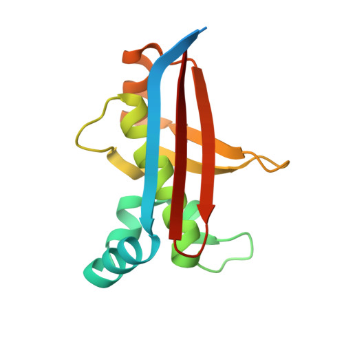
    The transfer of the phosphopantetheine chain from coenzyme A (CoA) to the acyl carrier protein (ACP), a key protein in both fatty acid and polyketide synthesis, is catalyzed by ACP synthase (AcpS). Streptomyces coelicolor AcpS is a doubly promiscuous enzyme capable of activation of ACPs from both fatty acid and polyketide synthesis and catalyzes the transfer of modified CoA substrates. Five crystal structures have been determined, including those of ligand-free AcpS, complexes with CoA and acetyl-CoA, and two of the active site mutants, His110Ala and Asp111Ala. All five structures are trimeric and provide further insight into the mechanism of catalysis, revealing the first detailed structure of a group I active site with the essential magnesium in place. Modeling of ACP binding supported by mutational analysis suggests an explanation for the promiscuity in terms of both ACP partner and modified CoA substrates.


  • Organizational Affiliation
    • Department of Biochemistry, School of Medical Sciences, University of Bristol, University Walk, Clifton, Bristol BS8 1TD, U.K.

Macromolecule Content 

  • Total Structure Weight: 44.66 kDa 
  • Atom Count: 2,924 
  • Modeled Residue Count: 369 
  • Deposited Residue Count: 429 
  • Unique protein chains: 1

Macromolecules

Find similar proteins by:|  3D Structure
Entity ID: 1
MoleculeChains  Sequence LengthOrganismDetailsImage
HOLO-[ACYL-CARRIER-PROTEIN] SYNTHASE
A, B, C
143Streptomyces coelicolorMutation(s): 0 
EC: 2.7.8.7
UniProt
Find proteins for O86785 (Streptomyces coelicolor (strain ATCC BAA-471 / A3(2) / M145))
Explore O86785 
Go to UniProtKB:  O86785
Entity Groups
Sequence Clusters30% Identity50% Identity70% Identity90% Identity95% Identity100% Identity
UniProt GroupO86785
Sequence Annotations
Expand
Reference Sequence

Experimental Data & Validation

Experimental Data

  • Method: X-RAY DIFFRACTION
  • Resolution: 1.98 Å
  • R-Value Free:  0.220 (Depositor), 0.228 (DCC) 
  • R-Value Work:  0.178 (Depositor), 0.182 (DCC) 
  • R-Value Observed: 0.180 (Depositor) 
Space Group: P 31 2 1
Unit Cell:
Length ( Å )Angle ( ˚ )
a = 75.94α = 90
b = 75.94β = 90
c = 108.351γ = 120
Software Package:
Software NamePurpose
REFMACrefinement

Structure Validation

View Full Validation Report


Entry History 

Deposition Data

Revision History  (Full details and data files)

  • Version 1.0: 2007-01-30
    Type: Initial release
  • Version 1.1: 2011-11-16
    Changes: Database references, Derived calculations, Non-polymer description, Other, Version format compliance
  • Version 1.2: 2023-12-13
    Changes: Data collection, Database references, Derived calculations, Other, Refinement description