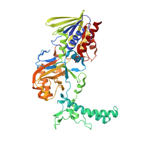
The Structure of a Bacterial L-Amino Acid Oxidase from Rhodococcus Opacus Gives New Evidence for the Hydride Mechanism for Dehydrogenation
Faust, A., Niefind, K., Hummel, W., Schomburg, D.(2007) J Mol Biology 367: 234
- PubMed: 17234209 Search on PubMed
- DOI: https://doi.org/10.1016/j.jmb.2006.11.071
- Primary Citation Related Structures:
2JAE, 2JB1, 2JB2, 2JB3 - PubMed Abstract:
l-Amino acid oxidase from Rhodococcus opacus (roLAAO) is classified as a member of the GR(2)-family of flavin-dependent oxidoreductases according to a highly conserved sequence motif for the cofactor binding. The monomer of the homodimeric enzyme consists of three well-defined domains: the FAD-binding domain corresponding to a general topology throughout the whole GR(2)-family; a substrate-binding domain with almost the same topology as the snake venom LAAO and a helical domain exclusively responsible for the unusual dimerisation mode of the enzyme and not found in other members of the family so far. We describe here high-resolution structures of the binary complex of protein and cofactor as well as the ternary complexes of protein, cofactor and ligands. This structures in addition to the structural knowledge of snake venom LAAO and DAAO from yeast and pig kidney permit more insight into different steps in the reaction mechanism of this class of enzymes. There is strong evidence for hydride transfer as the mechanism of dehydrogenation. This mechanism appears to be uncommon in a sense that the chemical transformation can proceed efficiently without the involvement of amino acid functional groups. Most groups present at the active site are involved in substrate recognition, binding and fixation, i.e. they direct the trajectory of the interacting orbitals. In this mode of catalysis orbital steering/interactions are the predominant factors for the chemical step(s). A mirror-symmetrical relationship between the two substrate-binding sites of d and l-amino acid oxidases is observed which facilitates enantiomeric selectivity while preserving a common arrangement of the residues in the active site. These results are of general relevance for the mechanism of flavoproteins and lead to the proposal of a common dehydrogenation step in the mechanism for l and d-amino acid oxidases.
- Universität zu Köln, Institut für Biochemie, Zülpicher Strasse 47, D-50674 Köln, Germany.
Organizational Affiliation: