2J4R | pdb_00002j4r

Structural Study of the Aquifex aeolicus PPX-GPPA enzyme


Experimental Data Snapshot

  • Method: X-RAY DIFFRACTION
  • Resolution: 2.71 Å
  • R-Value Free: 
    0.296 (Depositor), 0.218 (DCC) 
  • R-Value Work: 
    0.212 (Depositor), 0.209 (DCC) 
  • R-Value Observed: 
    0.212 (Depositor) 

Starting Model: experimental
View more details

wwPDB Validation 3D Report Full Report

Validation slider image for 2J4R

Ligand Structure Quality Assessment 


This is version 1.4 of the entry. See complete history

Literature

Structure of the Ppx/Gppa Phosphatase from Aquifex Aeolicus in Complex with the Alarmone Ppgpp

Kristensen, O.Ross, B.Gajhede, M.

(2008) J Mol Biology 375: 1469

  • DOI: https://doi.org/10.1016/j.jmb.2007.11.073
  • Primary Citation Related Structures: 
    2J4R

  • PubMed Abstract: 

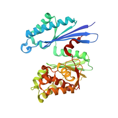
    The crystal structure of the prototype exopolyphosphatase/guanosine pentaphosphate phosphohydrolase protein family member from Aquifex aeolicus in complex with the intracellular second messenger guanosine tetraphosphate was determined at 2.7-A resolution. The hydrolytic base is identified as E119. The dual specificity established for the Escherichia coli homolog is shown to be compatible with a common active site for guanosine pentaphosphate and polyphosphate hydrolysis. Distinct and different degrees of closure between the two domains of the enzyme are associated with substrate binding. The arginines R22 and R267, residing in different domains, are crucial for guanosine pentaphosphate specificity as they interact with the unique 3'-ribose phosphorylation.


  • Organizational Affiliation
    • Biostructural Research, Department of Medicinal Chemistry, University of Copenhagen, Universitetsparken 2, DK-2100 Copenhagen, Denmark. ok@farma.ku.dk

Macromolecule Content 

  • Total Structure Weight: 70.6 kDa 
  • Atom Count: 4,997 
  • Modeled Residue Count: 600 
  • Deposited Residue Count: 616 
  • Unique protein chains: 1

Macromolecules

Find similar proteins by:|  3D Structure
Entity ID: 1
MoleculeChains  Sequence LengthOrganismDetailsImage
EXOPOLYPHOSPHATASE
A, B
308Aquifex aeolicusMutation(s): 1 
UniProt
Find proteins for O67040 (Aquifex aeolicus (strain VF5))
Explore O67040 
Go to UniProtKB:  O67040
Entity Groups
Sequence Clusters30% Identity50% Identity70% Identity90% Identity95% Identity100% Identity
UniProt GroupO67040
Sequence Annotations
Expand
Reference Sequence

Small Molecules

Ligands 1 Unique
IDChains Name / Formula / InChI Key2D Diagram3D Interactions
G4P

Query on G4P


Download:Ideal Coordinates CCD File
C [auth A]GUANOSINE-5',3'-TETRAPHOSPHATE
C10 H17 N5 O17 P4
BUFLLCUFNHESEH-UUOKFMHZSA-N

Experimental Data & Validation

Experimental Data

  • Method: X-RAY DIFFRACTION
  • Resolution: 2.71 Å
  • R-Value Free:  0.296 (Depositor), 0.218 (DCC) 
  • R-Value Work:  0.212 (Depositor), 0.209 (DCC) 
  • R-Value Observed: 0.212 (Depositor) 
Space Group: P 32 2 1
Unit Cell:
Length ( Å )Angle ( ˚ )
a = 70.46α = 90
b = 70.46β = 90
c = 223.71γ = 120
Software Package:
Software NamePurpose
CNSrefinement
XDSdata reduction

Structure Validation

View Full Validation Report


Ligand Structure Quality Assessment 


Entry History 

Deposition Data

Revision History  (Full details and data files)

  • Version 1.0: 2007-10-16
    Type: Initial release
  • Version 1.1: 2011-05-08
    Changes: Version format compliance
  • Version 1.2: 2011-07-13
    Changes: Version format compliance
  • Version 1.3: 2018-01-17
    Changes: Data collection
  • Version 1.4: 2023-12-13
    Changes: Data collection, Database references, Other, Refinement description, Structure summary