Analysis of Protein Hydration in Ultra-High Resolution Structures of the Srp Gtpase Ffh
Ramirez, U.D., Freymann, D.M.(2006) Acta Crystallogr D Biol Crystallogr 62: 1520
- PubMed: 17139088 Search on PubMedSearch on PubMed Central
- DOI: https://doi.org/10.1107/S0907444906040807
- Primary Citation Related Structures:
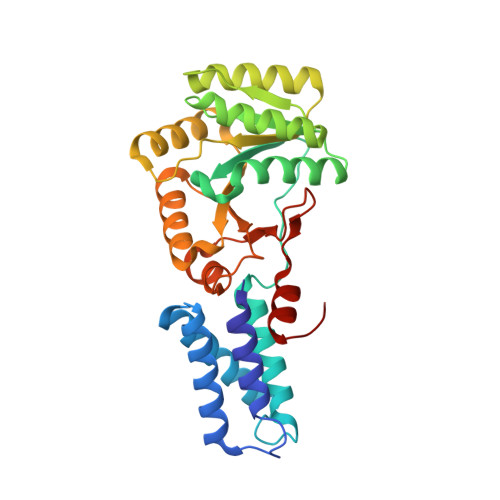
2J45, 2J46 - PubMed Abstract:
Two new structures of the SRP GTPase Ffh have been determined at 1.1 A resolution and provide the basis for comparative examination of the extensive water structure of the apo conformation of these GTPases. A set of well defined water-binding positions have been identified in the active site of the two-domain ;NG' GTPase, as well as at two functionally important interfaces. The water hydrogen-bonding network accommodates alternate conformations of the protein side chains by undergoing local rearrangements and, in one case, illustrates binding of a solute molecule within the active site by displacement of water molecules without further disruption of the water-interaction network. A subset of the water positions are well defined in several lower resolution structures, including those of different nucleotide-binding states; these appear to function in maintaining the protein structure. Consistent arrangements of surface water between three different ultrahigh-resolution structures provide a framework for beginning to understand how local water structure contributes to protein-ligand and protein-protein binding in the SRP GTPases.
- Department of Molecular Pharmacology and Biological Chemistry, Northwestern University, Chicago, IL 60611, USA.
Organizational Affiliation: