2IX7

Structure of apo-calmodulin bound to unconventional myosin V


Experimental Data Snapshot

  • Method: X-RAY DIFFRACTION
  • Resolution: 2.50 Å
  • R-Value Free: 0.259 
  • R-Value Work: 0.216 
  • R-Value Observed: 0.216 

wwPDB Validation   3D Report Full Report


This is version 1.2 of the entry. See complete history


Literature

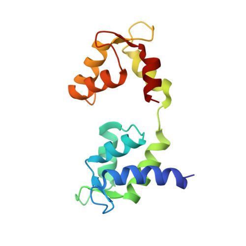
Crystal Structure of Apo-Calmodulin Bound to the First Two Iq Motifs of Myosin V Reveals Essential Recognition Features.

Houdusse, A.Gaucher, J.F.Krementsova, E.Mui, S.Trybus, K.M.Cohen, C.

(2006) Proc Natl Acad Sci U S A 103: 19326

  • DOI: 10.1073/pnas.0609436103
  • Primary Citation of Related Structures:  
    2IX7

  • PubMed Abstract: 
  • A 2.5-A resolution structure of calcium-free calmodulin (CaM) bound to the first two IQ motifs of the murine myosin V heavy chain reveals an unusual CaM conformation. The C-terminal lobe of each CaM adopts a semi-open conformation that grips the first part of the IQ motif (IQxxxR), whereas the N-terminal lobe adopts a closed conformation that interacts more weakly with the second part of the motif (GxxxR) ...

    A 2.5-A resolution structure of calcium-free calmodulin (CaM) bound to the first two IQ motifs of the murine myosin V heavy chain reveals an unusual CaM conformation. The C-terminal lobe of each CaM adopts a semi-open conformation that grips the first part of the IQ motif (IQxxxR), whereas the N-terminal lobe adopts a closed conformation that interacts more weakly with the second part of the motif (GxxxR). Variable residues in the IQ motif play a critical role in determining the precise structure of the bound CaM, such that even the consensus residues of different motifs show unique interactions with CaM. This complex serves as a model for the lever arm region of many classes of unconventional myosins, as well as other IQ motif-containing proteins such as neuromodulin and IQGAPs.


    Organizational Affiliation

    Motilité Structurale, Institut Curie, Centre National de la Recherche Scientifique, Unite Mixté de Recherche 144, 26 rue d'Ulm, 75248, Paris Cedex 05, France.


Macromolecules
Find similar proteins by:  (by identity cutoff)  |  3D Structure
Entity ID: 1
MoleculeChainsSequence LengthOrganismDetailsImage
CALMODULINA, B145Mus musculusMutation(s): 0 
Gene Names: Calm1CalmCamCam1
UniProt
Find proteins for P0DP26 (Mus musculus)
Explore P0DP26 
Go to UniProtKB:  P0DP26
Entity Groups  
Sequence Clusters30% Identity50% Identity70% Identity90% Identity95% Identity100% Identity
UniProt GroupP0DP26
Protein Feature View
Expand
  • Reference Sequence
Find similar proteins by:  (by identity cutoff)  |  3D Structure
Entity ID: 2
MoleculeChainsSequence LengthOrganismDetailsImage
MYOSIN-5AC58Mus musculusMutation(s): 0 
Gene Names: Myo5aDilute
UniProt & NIH Common Fund Data Resources
Find proteins for Q99104 (Mus musculus)
Explore Q99104 
Go to UniProtKB:  Q99104
IMPC:  MGI:105976
Entity Groups  
Sequence Clusters30% Identity50% Identity70% Identity90% Identity95% Identity100% Identity
UniProt GroupQ99104
Protein Feature View
Expand
  • Reference Sequence
Experimental Data & Validation

Experimental Data

  • Method: X-RAY DIFFRACTION
  • Resolution: 2.50 Å
  • R-Value Free: 0.259 
  • R-Value Work: 0.216 
  • R-Value Observed: 0.216 
  • Space Group: P 32 2 1
Unit Cell:
Length ( Å )Angle ( ˚ )
a = 112α = 90
b = 112β = 90
c = 102.34γ = 120
Software Package:
Software NamePurpose
CNSrefinement
MOSFLMdata reduction
SCALEPACKdata scaling
MLPHAREphasing

Structure Validation

View Full Validation Report


Entry History 

Deposition Data

Revision History  (Full details and data files)

  • Version 1.0: 2006-12-13
    Type: Initial release
  • Version 1.1: 2013-12-04
    Changes: Data collection, Derived calculations, Non-polymer description, Other, Refinement description, Source and taxonomy, Version format compliance
  • Version 1.2: 2019-02-27
    Changes: Data collection, Derived calculations, Experimental preparation, Other