2IUW | pdb_00002iuw

Crystal structure of human ABH3 in complex with iron ion and 2- oxoglutarate


Experimental Data Snapshot

  • Method: X-RAY DIFFRACTION
  • Resolution: 1.50 Å
  • R-Value Free: 
    0.191 (Depositor), 0.174 (DCC) 
  • R-Value Work: 
    0.131 (DCC) 
  • R-Value Observed: 
    0.141 (Depositor) 

wwPDB Validation 3D Report Full Report

Validation slider image for 2IUW

This is version 1.4 of the entry. See complete history

Literature

Human Abh3 Structure and Key Residues for Oxidative Demethylation to Reverse DNA/RNA Damage.

Sundheim, O.Vagbo, C.B.Bjoras, M.Desousa, M.M.L.Talstad, V.Aas, P.A.Drablos, F.Krokan, H.E.Tainer, J.A.Slupphaug, G.

(2006) EMBO J 25: 3389

  • DOI: https://doi.org/10.1038/sj.emboj.7601219
  • Primary Citation Related Structures: 
    2IUW

  • PubMed Abstract: 

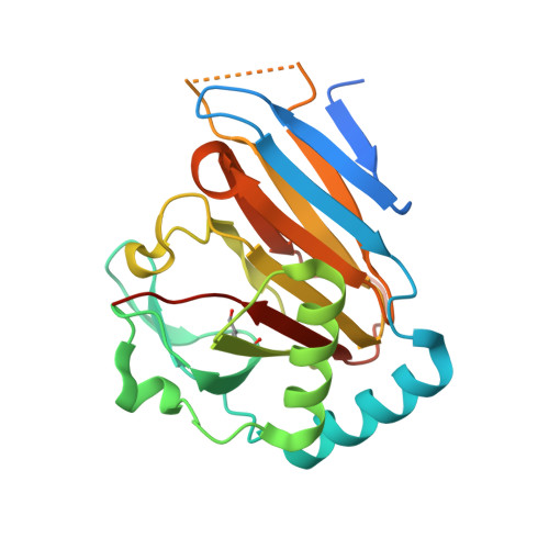
    Methylating agents are ubiquitous in the environment, and central in cancer therapy. The 1-methyladenine and 3-methylcytosine lesions in DNA/RNA contribute to the cytotoxicity of such agents. These lesions are directly reversed by ABH3 (hABH3) in humans and AlkB in Escherichia coli. Here, we report the structure of the hABH3 catalytic core in complex with iron and 2-oxoglutarate (2OG) at 1.5 A resolution and analyse key site-directed mutants. The hABH3 structure reveals the beta-strand jelly-roll fold that coordinates a catalytically active iron centre by a conserved His1-X-Asp/Glu-X(n)-His2 motif. This experimentally establishes hABH3 as a structural member of the Fe(II)/2OG-dependent dioxygenase superfamily, which couples substrate oxidation to conversion of 2OG into succinate and CO2. A positively charged DNA/RNA binding groove indicates a distinct nucleic acid binding conformation different from that predicted in the AlkB structure with three nucleotides. These results uncover previously unassigned key catalytic residues, identify a flexible hairpin involved in nucleotide flipping and ss/ds-DNA discrimination, and reveal self-hydroxylation of an active site leucine that may protect against uncoupled generation of dangerous oxygen radicals.


  • Organizational Affiliation
    • Department of Cancer Research and Molecular Medicine, NTNU, Trondheim, Norway.

Macromolecule Content 

  • Total Structure Weight: 28.38 kDa 
  • Atom Count: 1,959 
  • Modeled Residue Count: 205 
  • Deposited Residue Count: 238 
  • Unique protein chains: 1

Macromolecules

Find similar proteins by:|  3D Structure
Entity ID: 1
MoleculeChains  Sequence LengthOrganismDetailsImage
ALKYLATED REPAIR PROTEIN ALKB HOMOLOG 3238Homo sapiensMutation(s): 0 
EC: 1.14.11 (PDB Primary Data), 1.14.11.33 (UniProt), 1.14.11.54 (UniProt)
UniProt & NIH Common Fund Data Resources
Find proteins for Q96Q83 (Homo sapiens)
Explore Q96Q83 
Go to UniProtKB:  Q96Q83
PHAROS:  Q96Q83
GTEx:  ENSG00000166199 
Entity Groups
Sequence Clusters30% Identity50% Identity70% Identity90% Identity95% Identity100% Identity
UniProt GroupQ96Q83
Sequence Annotations
Expand
Reference Sequence

Small Molecules

Ligands 3 Unique
IDChains Name / Formula / InChI Key2D Diagram3D Interactions
AKG

Query on AKG


Download:Ideal Coordinates CCD File
E [auth A]2-OXOGLUTARIC ACID
C5 H6 O5
KPGXRSRHYNQIFN-UHFFFAOYSA-N
BME

Query on BME


Download:Ideal Coordinates CCD File
F [auth A],
G [auth A],
H [auth A],
I [auth A]
BETA-MERCAPTOETHANOL
C2 H6 O S
DGVVWUTYPXICAM-UHFFFAOYSA-N
FE

Query on FE


Download:Ideal Coordinates CCD File
B [auth A],
C [auth A],
D [auth A]
FE (III) ION
Fe
VTLYFUHAOXGGBS-UHFFFAOYSA-N
Modified Residues  1 Unique
IDChains TypeFormula2D DiagramParent
LED
Query on LED
A
L-PEPTIDE LINKINGC6 H11 N O3LEU

Experimental Data & Validation

Experimental Data

  • Method: X-RAY DIFFRACTION
  • Resolution: 1.50 Å
  • R-Value Free:  0.191 (Depositor), 0.174 (DCC) 
  • R-Value Work:  0.131 (DCC) 
  • R-Value Observed: 0.141 (Depositor) 
Space Group: I 41
Unit Cell:
Length ( Å )Angle ( ˚ )
a = 119.955α = 90
b = 119.955β = 90
c = 40.554γ = 90
Software Package:
Software NamePurpose
SHELXL-97refinement
HKL-2000data reduction
HKL-2000data scaling
SOLVEphasing

Structure Validation

View Full Validation Report


Entry History 

Deposition Data

Revision History  (Full details and data files)

  • Version 1.0: 2006-07-26
    Type: Initial release
  • Version 1.1: 2011-05-08
    Changes: Version format compliance
  • Version 1.2: 2011-07-13
    Changes: Version format compliance
  • Version 1.3: 2019-05-22
    Changes: Data collection, Derived calculations, Other, Refinement description
  • Version 1.4: 2025-04-09
    Changes: Data collection, Database references, Derived calculations, Other, Structure summary